• 文献检索
  • 文档翻译
  • 深度研究
  • 学术资讯
  • Suppr Zotero 插件Zotero 插件
  • 邀请有礼
  • 套餐&价格
  • 历史记录
应用&插件
Suppr Zotero 插件Zotero 插件浏览器插件Mac 客户端Windows 客户端微信小程序
定价
高级版会员购买积分包购买API积分包
服务
文献检索文档翻译深度研究API 文档MCP 服务
关于我们
关于 Suppr公司介绍联系我们用户协议隐私条款
关注我们

Suppr 超能文献

核心技术专利:CN118964589B侵权必究
粤ICP备2023148730 号-1Suppr @ 2026

文献检索

告别复杂PubMed语法,用中文像聊天一样搜索,搜遍4000万医学文献。AI智能推荐,让科研检索更轻松。

立即免费搜索

文件翻译

保留排版,准确专业,支持PDF/Word/PPT等文件格式,支持 12+语言互译。

免费翻译文档

深度研究

AI帮你快速写综述,25分钟生成高质量综述,智能提取关键信息,辅助科研写作。

立即免费体验

早期胚胎中染色质接触高分辨率作图揭示的拓扑结构域边界、绝缘体和多线染色体间带的收敛

Convergence of topological domain boundaries, insulators, and polytene interbands revealed by high-resolution mapping of chromatin contacts in the early embryo.

机构信息

Department of Molecular and Cell Biology, University of California, Berkeley, CA, United States.

Department of Integrative Biology, University of California, Berkeley, CA, United States.

出版信息

Elife. 2017 Nov 17;6:e29550. doi: 10.7554/eLife.29550.

DOI:10.7554/eLife.29550
PMID:29148971
原文链接:https://pmc.ncbi.nlm.nih.gov/articles/PMC5739541/
Abstract

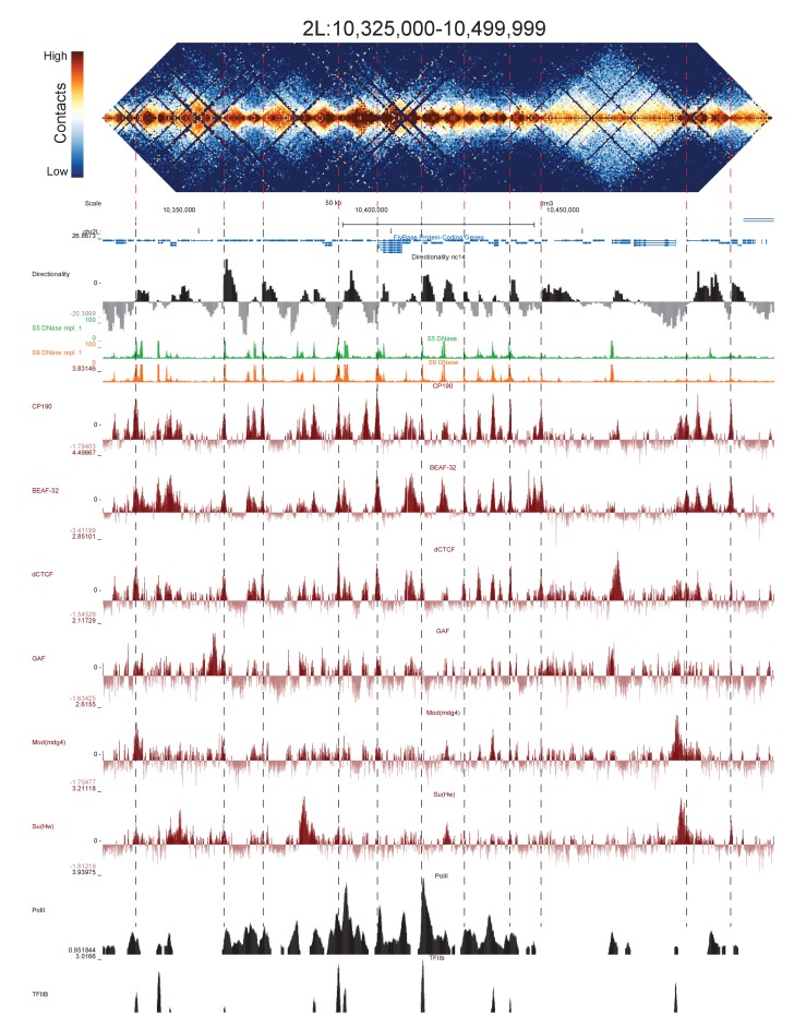
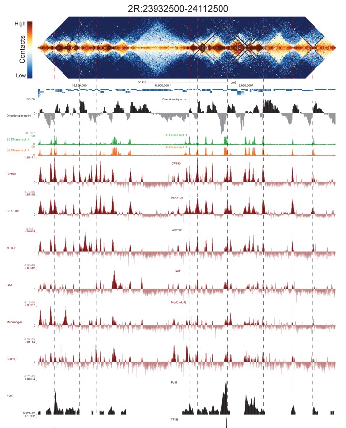
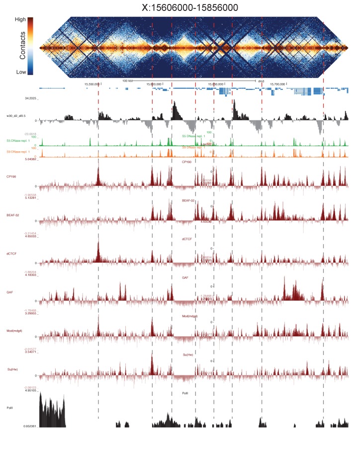
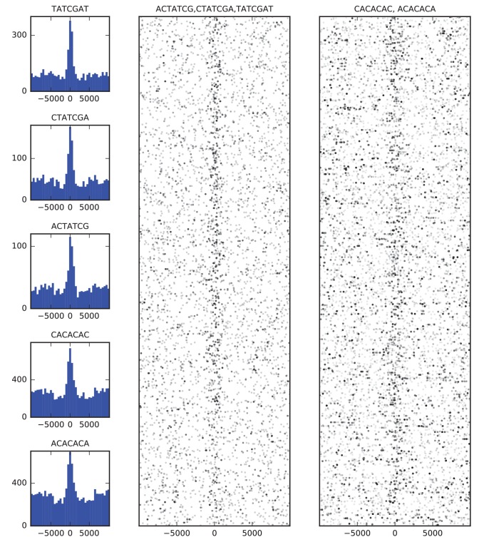
High-throughput assays of three-dimensional interactions of chromosomes have shed considerable light on the structure of animal chromatin. Despite this progress, the precise physical nature of observed structures and the forces that govern their establishment remain poorly understood. Here we present high resolution Hi-C data from early embryos. We demonstrate that boundaries between topological domains of various sizes map to DNA elements that resemble classical insulator elements: short genomic regions sensitive to DNase digestion that are strongly bound by known insulator proteins and are frequently located between divergent promoters. Further, we show a striking correspondence between these elements and the locations of mapped polytene interband regions. We believe it is likely this relationship between insulators, topological boundaries, and polytene interbands extends across the genome, and we therefore propose a model in which decompaction of boundary-insulator-interband regions drives the organization of interphase chromosomes by creating stable physical separation between adjacent domains.

摘要

高通量分析染色体的三维相互作用为动物染色质的结构提供了重要线索。尽管取得了这些进展,但观察到的结构的精确物理性质以及控制其形成的力仍未得到很好的理解。在这里,我们展示了来自早期胚胎的高分辨率 Hi-C 数据。我们证明,不同大小的拓扑域之间的边界映射到类似于经典绝缘子元件的 DNA 元件:对 DNA 酶消化敏感的短基因组区域,由已知的绝缘子蛋白强烈结合,并且经常位于发散启动子之间。此外,我们还展示了这些元件与映射的多线染色体间带区的位置之间存在惊人的对应关系。我们认为,绝缘子、拓扑边界和多线染色体间带之间的这种关系很可能在整个基因组中延伸,因此我们提出了一个模型,即边界-绝缘子-间带区域的解压缩通过在相邻域之间建立稳定的物理分离来驱动间期染色体的组织。

https://cdn.ncbi.nlm.nih.gov/pmc/blobs/b631/5739541/af101cad223d/elife-29550-fig9.jpg
https://cdn.ncbi.nlm.nih.gov/pmc/blobs/b631/5739541/0c7e6a67763a/elife-29550-fig1.jpg
https://cdn.ncbi.nlm.nih.gov/pmc/blobs/b631/5739541/f4f814eac69c/elife-29550-fig2.jpg
https://cdn.ncbi.nlm.nih.gov/pmc/blobs/b631/5739541/8762877093a4/elife-29550-fig2-figsupp1.jpg
https://cdn.ncbi.nlm.nih.gov/pmc/blobs/b631/5739541/050f57a3873e/elife-29550-fig2-figsupp2.jpg
https://cdn.ncbi.nlm.nih.gov/pmc/blobs/b631/5739541/7b2fa64f9ade/elife-29550-fig2-figsupp3.jpg
https://cdn.ncbi.nlm.nih.gov/pmc/blobs/b631/5739541/f005b767012b/elife-29550-fig2-figsupp4.jpg
https://cdn.ncbi.nlm.nih.gov/pmc/blobs/b631/5739541/1ea85938826d/elife-29550-fig2-figsupp5.jpg
https://cdn.ncbi.nlm.nih.gov/pmc/blobs/b631/5739541/06331a63576c/elife-29550-fig3.jpg
https://cdn.ncbi.nlm.nih.gov/pmc/blobs/b631/5739541/3effd88dedbc/elife-29550-fig3-figsupp1.jpg
https://cdn.ncbi.nlm.nih.gov/pmc/blobs/b631/5739541/ac5874a8ffc6/elife-29550-fig3-figsupp2.jpg
https://cdn.ncbi.nlm.nih.gov/pmc/blobs/b631/5739541/ca357e12c10e/elife-29550-fig3-figsupp3.jpg
https://cdn.ncbi.nlm.nih.gov/pmc/blobs/b631/5739541/a3e0090bee45/elife-29550-fig3-figsupp4.jpg
https://cdn.ncbi.nlm.nih.gov/pmc/blobs/b631/5739541/b91000ae6048/elife-29550-fig4.jpg
https://cdn.ncbi.nlm.nih.gov/pmc/blobs/b631/5739541/7e7f4e8fe0f0/elife-29550-fig5.jpg
https://cdn.ncbi.nlm.nih.gov/pmc/blobs/b631/5739541/47acd9cd2da6/elife-29550-fig5-figsupp1.jpg
https://cdn.ncbi.nlm.nih.gov/pmc/blobs/b631/5739541/4a6424b3e415/elife-29550-fig5-figsupp2.jpg
https://cdn.ncbi.nlm.nih.gov/pmc/blobs/b631/5739541/dbebabb33cdb/elife-29550-fig6.jpg
https://cdn.ncbi.nlm.nih.gov/pmc/blobs/b631/5739541/99de80b1551f/elife-29550-fig7.jpg
https://cdn.ncbi.nlm.nih.gov/pmc/blobs/b631/5739541/2a321848655f/elife-29550-fig7-figsupp1.jpg
https://cdn.ncbi.nlm.nih.gov/pmc/blobs/b631/5739541/ec0747e61c87/elife-29550-fig8.jpg
https://cdn.ncbi.nlm.nih.gov/pmc/blobs/b631/5739541/02566cd98899/elife-29550-fig8-figsupp1.jpg
https://cdn.ncbi.nlm.nih.gov/pmc/blobs/b631/5739541/2e0220ba8d2e/elife-29550-fig8-figsupp2.jpg
https://cdn.ncbi.nlm.nih.gov/pmc/blobs/b631/5739541/ba4cb946b0a2/elife-29550-fig8-figsupp3.jpg
https://cdn.ncbi.nlm.nih.gov/pmc/blobs/b631/5739541/6c811f58255c/elife-29550-fig8-figsupp4.jpg
https://cdn.ncbi.nlm.nih.gov/pmc/blobs/b631/5739541/2e0740866b85/elife-29550-fig8-figsupp5.jpg
https://cdn.ncbi.nlm.nih.gov/pmc/blobs/b631/5739541/f5760c8c994f/elife-29550-fig8-figsupp6.jpg
https://cdn.ncbi.nlm.nih.gov/pmc/blobs/b631/5739541/086f0d93e15e/elife-29550-fig8-figsupp7.jpg
https://cdn.ncbi.nlm.nih.gov/pmc/blobs/b631/5739541/660d3d88d7b7/elife-29550-fig8-figsupp8.jpg
https://cdn.ncbi.nlm.nih.gov/pmc/blobs/b631/5739541/1be5222bf6d8/elife-29550-fig8-figsupp9.jpg
https://cdn.ncbi.nlm.nih.gov/pmc/blobs/b631/5739541/764c950919e2/elife-29550-fig8-figsupp10.jpg
https://cdn.ncbi.nlm.nih.gov/pmc/blobs/b631/5739541/af101cad223d/elife-29550-fig9.jpg

相似文献

1
Convergence of topological domain boundaries, insulators, and polytene interbands revealed by high-resolution mapping of chromatin contacts in the early embryo.早期胚胎中染色质接触高分辨率作图揭示的拓扑结构域边界、绝缘体和多线染色体间带的收敛
Elife. 2017 Nov 17;6:e29550. doi: 10.7554/eLife.29550.
2
Banding Pattern of Polytene Chromosomes as a Representation of Universal Principles of Chromatin Organization into Topological Domains.多线染色体的带型作为染色质组织成拓扑结构域的普遍原则的一种体现。
Biochemistry (Mosc). 2018 Apr;83(4):338-349. doi: 10.1134/S0006297918040053.
3
[Chromomeric organization of interphase chromosomes in Drosophila melanogaster].[黑腹果蝇间期染色体的染色粒组织]
Tsitologiia. 2013;55(3):144-7.
4
High-resolution in situ hybridization analysis on the chromosomal interval 61C7-61C8 of Drosophila melanogaster reveals interbands as open chromatin domains.对黑腹果蝇61C7 - 61C8染色体区间进行的高分辨率原位杂交分析表明,间带是开放染色质结构域。
Chromosoma. 2016 Jun;125(3):423-35. doi: 10.1007/s00412-015-0554-5. Epub 2015 Oct 31.
5
Genes Containing Long Introns Occupy Series of Bands and Interbands In polytene Chromosomes.含有长内含子的基因占据多线染色体的一系列带和带间区。
Genes (Basel). 2020 Apr 11;11(4):417. doi: 10.3390/genes11040417.
6
Genetic organization of interphase chromosome bands and interbands in Drosophila melanogaster.黑腹果蝇间期染色体带和间带的遗传组织
PLoS One. 2014 Jul 29;9(7):e101631. doi: 10.1371/journal.pone.0101631. eCollection 2014.
7
ECTOPIC TETHERING OF THE CHROMATOR PROTEIN IN UASDBD(GAL4) SYSTEM AS APPROACH FOR STUDYING OF THE INSULATOR PROTEINS IN DROSOPHILA MELANOGASTER POLYTENE CHROMOSOMES.在UASDBD(GAL4)系统中对染色质蛋白进行异位束缚作为研究黑腹果蝇多线染色体中绝缘子蛋白的方法。
Tsitologiia. 2016;58(6):493-7.
8
Phosphorylated histone variant γH2Av is associated with chromatin insulators in Drosophila.磷酸化组蛋白变体 γH2Av 与果蝇中的染色质绝缘子有关。
PLoS Genet. 2022 Oct 5;18(10):e1010396. doi: 10.1371/journal.pgen.1010396. eCollection 2022 Oct.
9
Architecture of Promoters of House-Keeping Genes in Polytene Chromosome Interbands of Drosophila melanogaster.黑腹果蝇多线染色体间带中管家基因启动子的结构
Dokl Biochem Biophys. 2019 Mar;485(1):95-100. doi: 10.1134/S1607672919020029. Epub 2019 Jun 14.
10
LOCALIZATION OF MOBILE P-ELEMENT INSERTIONS IN DOMAINS OF ACTIVE AND REPRESSED CHROMATIN IN DROSOPHILA MELANOGASTER’S GENOME.黑腹果蝇基因组中活性和抑制性染色质区域内移动P因子插入的定位
Tsitologiia. 2016;58(4):258-61.

引用本文的文献

1
Stem-loop and circle-loop TADs generated by directional pairing of boundary elements have distinct physical and regulatory properties.由边界元件定向配对产生的茎环和环loop TADs 具有不同的物理和调控性质。
Elife. 2024 Aug 7;13:RP94114. doi: 10.7554/eLife.94114.
2
Genome organization regulates nuclear pore complex formation and promotes differentiation during oogenesis.基因组组织调控核孔复合体的形成并促进卵母细胞发生中的分化。
Genes Dev. 2024 Jun 25;38(9-10):436-454. doi: 10.1101/gad.351402.123.
3
The probability of chromatin to be at the nuclear lamina has no systematic effect on its transcription level in fruit flies.

本文引用的文献

1
High-resolution TADs reveal DNA sequences underlying genome organization in flies.高分辨率拓扑关联结构域揭示果蝇基因组组织背后的DNA序列。
Nat Commun. 2018 Jan 15;9(1):189. doi: 10.1038/s41467-017-02525-w.
2
Chromatin Architecture Emerges during Zygotic Genome Activation Independent of Transcription.染色质结构在合子基因组激活过程中独立于转录而出现。
Cell. 2017 Apr 6;169(2):216-228.e19. doi: 10.1016/j.cell.2017.03.024.
3
Complex multi-enhancer contacts captured by genome architecture mapping.通过基因组结构图谱捕获的复杂多增强子接触。
在果蝇中,染色质位于核层的概率与其转录水平没有系统的关系。
Epigenetics Chromatin. 2024 May 6;17(1):13. doi: 10.1186/s13072-024-00528-8.
4
Developmental and Housekeeping Genes: Two Types of Genetic Organization in the Genome.发育基因和管家基因:基因组中的两种遗传组织形式。
Int J Mol Sci. 2024 Apr 6;25(7):4068. doi: 10.3390/ijms25074068.
5
-regulatory modes of inactivation in butterfly forewings.蝴蝶前翅的失活调控模式。
Elife. 2024 Jan 23;12:RP90846. doi: 10.7554/eLife.90846.
6
Genome organization regulates nuclear pore complex formation and promotes differentiation during Drosophila oogenesis.基因组组织调控果蝇卵子发生过程中的核孔复合体形成并促进分化。
bioRxiv. 2023 Nov 16:2023.11.15.567233. doi: 10.1101/2023.11.15.567233.
7
Setting the stage for development: the maternal-to-zygotic transition in Drosophila.为发育奠定基础:果蝇中的母体到合子过渡。
Genetics. 2023 Oct 4;225(2). doi: 10.1093/genetics/iyad142.
8
Chromosome-level organization of the regulatory genome in the Drosophila nervous system.果蝇神经系统调控基因组的染色体水平组织。
Cell. 2023 Aug 31;186(18):3826-3844.e26. doi: 10.1016/j.cell.2023.07.008. Epub 2023 Aug 2.
9
Localization of the Drosophila pioneer factor GAF to subnuclear foci is driven by DNA binding and required to silence satellite repeat expression.果蝇先驱因子 GAF 的核内定位是由 DNA 结合驱动的,并且需要沉默卫星重复序列的表达。
Dev Cell. 2023 Sep 11;58(17):1610-1624.e8. doi: 10.1016/j.devcel.2023.06.010. Epub 2023 Jul 20.
10
Precisely timed regulation of enhancer activity defines the binary expression pattern of Fushi tarazu in the Drosophila embryo.精确调控增强子活性决定了 Fushi tarazu 在果蝇胚胎中的二元表达模式。
Curr Biol. 2023 Jul 24;33(14):2839-2850.e7. doi: 10.1016/j.cub.2023.04.005. Epub 2023 Apr 27.
Nature. 2017 Mar 23;543(7646):519-524. doi: 10.1038/nature21411. Epub 2017 Mar 8.
4
Computational Identification of Genomic Features That Influence 3D Chromatin Domain Formation.影响三维染色质结构域形成的基因组特征的计算识别
PLoS Comput Biol. 2016 May 20;12(5):e1004908. doi: 10.1371/journal.pcbi.1004908. eCollection 2016 May.
5
Stable Chromosome Condensation Revealed by Chromosome Conformation Capture.通过染色体构象捕获揭示的稳定染色体凝聚
Cell. 2015 Nov 5;163(4):934-46. doi: 10.1016/j.cell.2015.10.026.
6
Active chromatin and transcription play a key role in chromosome partitioning into topologically associating domains.活跃染色质和转录在染色体划分为拓扑相关结构域的过程中起关键作用。
Genome Res. 2016 Jan;26(1):70-84. doi: 10.1101/gr.196006.115. Epub 2015 Oct 30.
7
CRISPR Inversion of CTCF Sites Alters Genome Topology and Enhancer/Promoter Function.CTCF位点的CRISPR倒置改变基因组拓扑结构和增强子/启动子功能。
Cell. 2015 Aug 13;162(4):900-10. doi: 10.1016/j.cell.2015.07.038.
8
Condensin-driven remodelling of X chromosome topology during dosage compensation.剂量补偿过程中凝聚素驱动的X染色体拓扑结构重塑。
Nature. 2015 Jul 9;523(7559):240-4. doi: 10.1038/nature14450. Epub 2015 Jun 1.
9
Disruptions of topological chromatin domains cause pathogenic rewiring of gene-enhancer interactions.拓扑染色质结构域的破坏会导致基因-增强子相互作用的致病性重排。
Cell. 2015 May 21;161(5):1012-1025. doi: 10.1016/j.cell.2015.04.004. Epub 2015 May 7.
10
Widespread rearrangement of 3D chromatin organization underlies polycomb-mediated stress-induced silencing.3D染色质组织的广泛重排是多梳蛋白介导的应激诱导沉默的基础。
Mol Cell. 2015 Apr 16;58(2):216-31. doi: 10.1016/j.molcel.2015.02.023. Epub 2015 Mar 26.