Department of Biology, Temple University, Philadelphia, Pennsylvania, 19122, USA.
Department of Cell and Developmental Biology, University of Michigan Medical School, Ann Arbor, MI, 48109, USA.
Sci Rep. 2017 Nov 17;7(1):15793. doi: 10.1038/s41598-017-16103-z.
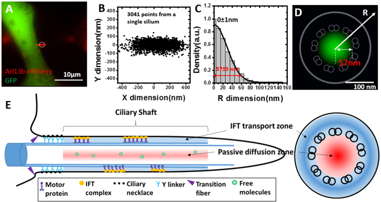
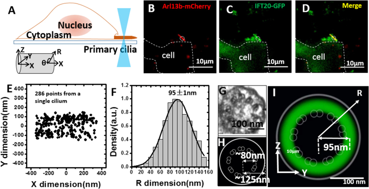
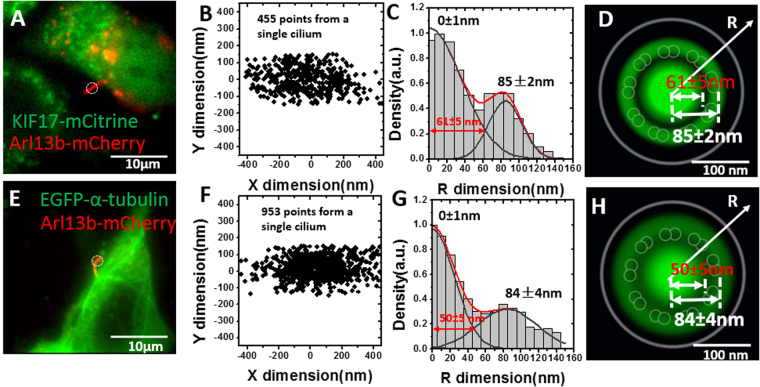
Transport of membrane and cytosolic proteins in primary cilia is thought to depend on intraflagellar transport (IFT) and diffusion. However, the relative contribution and spatial routes of each transport mechanism are largely unknown. Although challenging to decipher, the details of these routes are essential for our understanding of protein transport in primary cilia, a critically affected process in many genetic diseases. By using a high-speed virtual 3D super-resolution microscopy, we have mapped the 3D spatial locations of transport routes for various cytosolic proteins in the 250-nm-wide shaft of live primary cilia with a spatiotemporal resolution of 2 ms and <16 nm. Our data reveal two spatially distinguishable transport routes for cytosolic proteins: an IFT-dependent path along the axoneme, and a passive-diffusion route in the axonemal lumen that escaped previous studies. While all cytosolic proteins tested primarily utilize the IFT path in the anterograde direction, differences are observed in the retrograde direction where IFT20 only utilizes IFT, and approximately half of KIF17 and one third of α-tubulin utilizes diffusion besides IFT.
原发性纤毛内的膜蛋白和胞质蛋白的运输被认为依赖于鞭毛内运输(IFT)和扩散。然而,每种运输机制的相对贡献和空间途径在很大程度上尚不清楚。尽管这些途径的细节难以解读,但对于我们理解原发性纤毛中的蛋白运输至关重要,因为许多遗传疾病都受到该过程的严重影响。通过使用高速虚拟 3D 超分辨率显微镜,我们以 2 毫秒和 <16nm 的时空分辨率,绘制了活的原发性纤毛 250nm 宽的轴丝中各种胞质蛋白的运输途径的 3D 空间位置。我们的数据揭示了两种在空间上可区分的胞质蛋白运输途径:一种是沿着轴丝的 IFT 依赖途径,另一种是在轴丝腔中的被动扩散途径,这两种途径在先前的研究中都没有被发现。虽然所有测试的胞质蛋白在顺行方向上主要利用 IFT 途径,但在逆行方向上存在差异,其中 IFT20 仅利用 IFT,而大约一半的 KIF17 和三分之一的α-微管蛋白除了 IFT 之外还利用扩散。