Department of Radiation Oncology (MAASTRO), GROW - School for Oncology and Developmental Biology, Maastricht University Medical Center, Maastricht, the Netherlands.
Radiat Oncol. 2017 Nov 21;12(1):181. doi: 10.1186/s13014-017-0922-9.
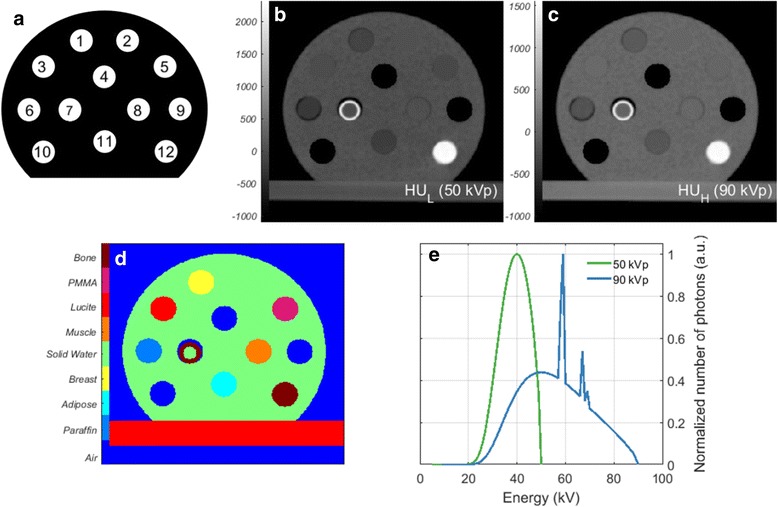
To investigate the feasibility of using dual-energy CT (DECT) for tissue segmentation and kilovolt (kV) dose calculations in pre-clinical studies and assess potential dose calculation accuracy gain.
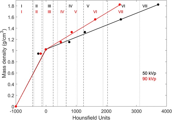
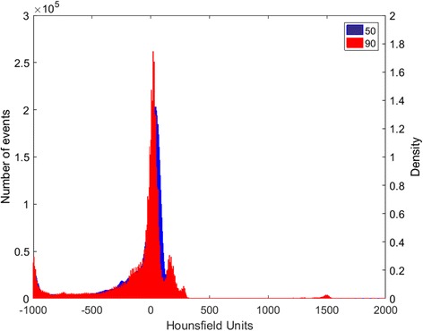
Two phantoms and an ex-vivo mouse were scanned in a small animal irradiator with two distinct energies. Tissue segmentation was performed with the single-energy CT (SECT) and DECT methods. A number of different material maps was used. Dose calculations were performed to verify the impact of segmentations on the dose accuracy.
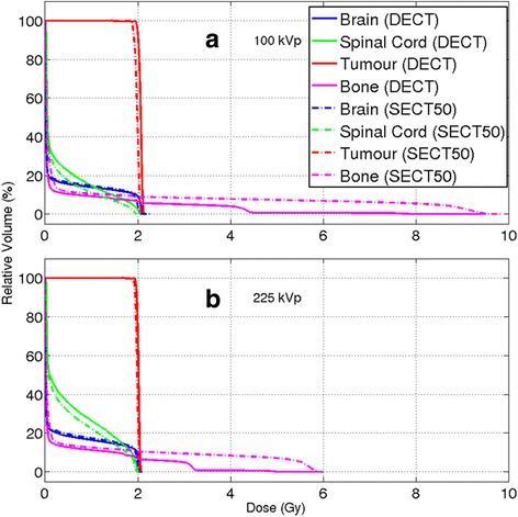
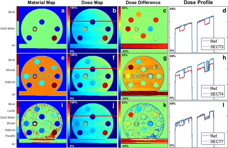
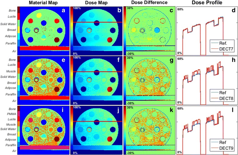
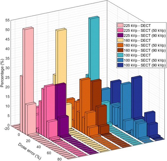
DECT showed better overall results in comparison to SECT. Higher number of DECT segmentation media resulted in smaller dose differences in comparison to the reference. Increasing the number of materials in the SECT method yielded more instability. Both modalities showed a limit to which adding more materials with similar characteristics ceased providing better segmentation results, and resulted in more noise in the material maps and the dose distributions. The effect was aggravated with a decrease in beam energy. For the ex-vivo specimen, the choice of only one high dense bone for the SECT method resulted in large volumes of tissue receiving high doses. For the DECT method, the choice of more than one kind of bone resulted in lower dose values for the different tissues occupying the same volume. For the organs at risk surrounded by bone, the doses were lower when using the SECT method in comparison to DECT, due to the high absorption of the bone. SECT material segmentation may lead to an underestimation of the dose to OAR in the proximity of bone.
The DECT method enabled the selection of a higher number of materials thereby increasing the accuracy in dose calculations. In phantom studies, SECT performed best with three materials and DECT with seven for the phantom case. For irradiations in preclinical studies with kV photon energies, the use of DECT segmentation combined with the choice of a low-density bone is recommended.
研究使用双能 CT(DECT)进行临床前研究中的组织分割和千伏(kV)剂量计算的可行性,并评估潜在的剂量计算准确性增益。
使用两种不同能量在小动物辐照器中对两个体模和一个离体小鼠进行扫描。使用单能量 CT(SECT)和 DECT 方法进行组织分割。使用了多种不同的材料图。进行剂量计算以验证分割对剂量准确性的影响。
DECT 与 SECT 相比显示出更好的整体结果。与参考值相比,更多的 DECT 分割介质导致剂量差异更小。在 SECT 方法中增加材料的数量会导致更多的不稳定性。两种方式都存在一个限制,即添加更多具有相似特征的材料不会提供更好的分割结果,并且会导致材料图和剂量分布中的噪声增加。随着束能的降低,这种影响会加剧。对于离体标本,SECT 方法仅选择一种高密度骨,导致大量组织接受高剂量。对于 DECT 方法,选择一种以上的骨导致不同组织占据相同体积的剂量值更低。对于被骨包围的危及器官,由于骨的高吸收率,使用 SECT 方法时剂量比 DECT 低。SECT 材料分割可能导致骨附近危及器官的剂量低估。
DECT 方法可以选择更多的材料,从而提高剂量计算的准确性。在体模研究中,SECT 在三种材料下表现最佳,而 DECT 在七种材料下表现最佳。对于临床前研究中的 kV 光子能量照射,建议使用 DECT 分割并选择低密度骨。