Malviya Manish, Barman Sumanta, Golombeck Kristin S, Planagumà Jesús, Mannara Francesco, Strutz-Seebohm Nathalie, Wrzos Claudia, Demir Fatih, Baksmeier Christine, Steckel Julia, Falk Kim Kristin, Gross Catharina C, Kovac Stjepana, Bönte Kathrin, Johnen Andreas, Wandinger Klaus-Peter, Martín-García Elena, Becker Albert J, Elger Christian E, Klöcker Nikolaj, Wiendl Heinz, Meuth Sven G, Hartung Hans-Peter, Seebohm Guiscard, Leypoldt Frank, Maldonado Rafael, Stadelmann Christine, Dalmau Josep, Melzer Nico, Goebels Norbert
Department of Neurology Medical Faculty Heinrich Heine University Düsseldorf Düsseldorf Germany.
Present address: Centre Physiopathologie de Toulouse-Purpan Université Toulouse III Toulouse France.
Ann Clin Transl Neurol. 2017 Oct 3;4(11):768-783. doi: 10.1002/acn3.444. eCollection 2017 Nov.
Autoimmune encephalitis is most frequently associated with anti-NMDAR autoantibodies. Their pathogenic relevance has been suggested by passive transfer of patients' cerebrospinal fluid (CSF) in mice in vivo. We aimed to analyze the intrathecal plasma cell repertoire, identify autoantibody-producing clones, and characterize their antibody signatures in recombinant form.
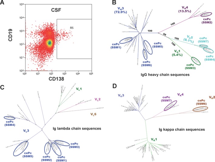
Patients with recent onset typical anti-NMDAR encephalitis were subjected to flow cytometry analysis of the peripheral and intrathecal immune response before, during, and after immunotherapy. Recombinant human monoclonal antibodies (rhuMab) were cloned and expressed from matching immunoglobulin heavy- (IgH) and light-chain (IgL) amplicons of clonally expanded intrathecal plasma cells (cePc) and tested for their pathogenic relevance.
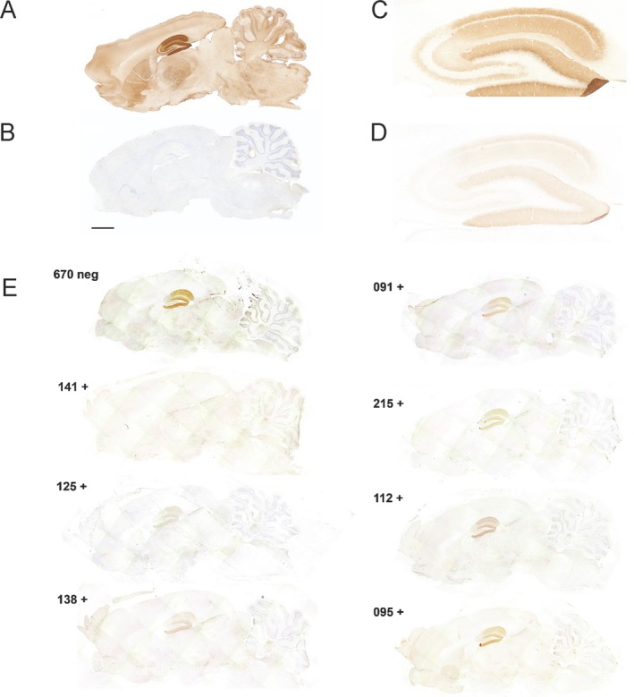
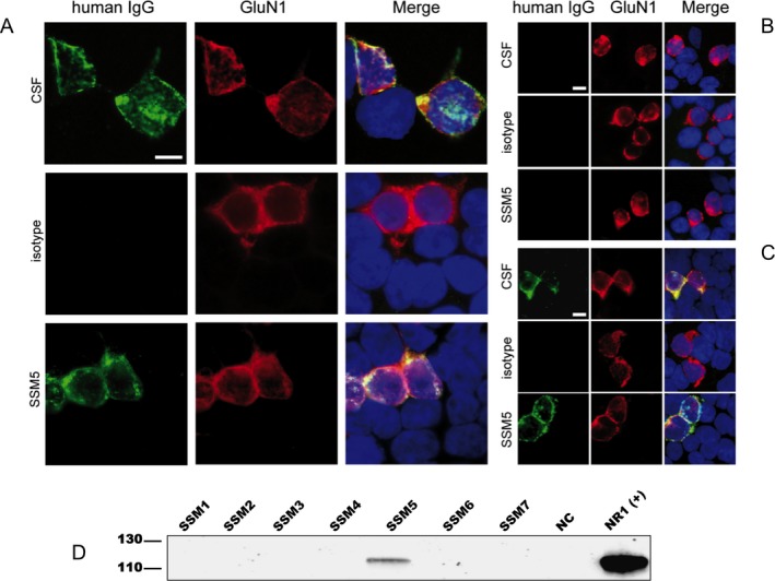
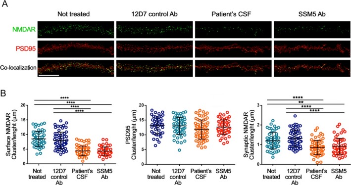
Intrathecal accumulation of B and plasma cells corresponded to the clinical course. The presence of cePc with hypermutated antigen receptors indicated an antigen-driven intrathecal immune response. Consistently, a single recombinant human GluN1-specific monoclonal antibody, rebuilt from intrathecal cePc, was sufficient to reproduce NMDAR epitope specificity in vitro. After intraventricular infusion in mice, it accumulated in the hippocampus, decreased synaptic NMDAR density, and caused severe reversible memory impairment, a key pathogenic feature of the human disease, in vivo.
A CNS-specific humoral immune response is present in anti-NMDAR encephalitis specifically targeting the GluN1 subunit of the NMDAR. Using reverse genetics, we recovered the typical intrathecal antibody signature in recombinant form, and proved its pathogenic relevance by passive transfer of disease symptoms from man to mouse, providing the critical link between intrathecal immune response and the pathogenesis of anti-NMDAR encephalitis as a humorally mediated autoimmune disease.
自身免疫性脑炎最常与抗NMDAR自身抗体相关。小鼠体内被动转移患者脑脊液(CSF)提示了它们的致病相关性。我们旨在分析鞘内浆细胞库,鉴定产生自身抗体的克隆,并以重组形式表征其抗体特征。
对近期发病的典型抗NMDAR脑炎患者在免疫治疗前、治疗期间和治疗后进行外周和鞘内免疫反应的流式细胞术分析。从克隆扩增的鞘内浆细胞(cePc)匹配的免疫球蛋白重链(IgH)和轻链(IgL)扩增子中克隆并表达重组人单克隆抗体(rhuMab),并测试其致病相关性。
B细胞和浆细胞的鞘内积聚与临床病程一致。具有高度突变抗原受体的cePc的存在表明存在抗原驱动的鞘内免疫反应。同样,从鞘内cePc重建的单一重组人GluN1特异性单克隆抗体足以在体外重现NMDAR表位特异性。在小鼠脑室内注入后,它在海马中积聚,降低突触NMDAR密度,并在体内引起严重的可逆性记忆障碍,这是人类疾病的关键致病特征。
抗NMDAR脑炎中存在针对NMDAR的GluN1亚基的中枢神经系统特异性体液免疫反应。通过反向遗传学,我们以重组形式恢复了典型的鞘内抗体特征,并通过将疾病症状从人被动转移到小鼠身上证明了其致病相关性,提供了鞘内免疫反应与作为体液介导的自身免疫性疾病的抗NMDAR脑炎发病机制之间的关键联系。