Tang Yan, Xiao Xue, Xie Hua, Wan Chang-Min, Meng Li, Liu Zhen-Hua, Liao Wei-Hua, Tang Bei-Sha, Guo Ji-Feng
Department of Neurology, Xiangya Hospital, Central South University, Changsha, China.
School of Information Science and Engineering, Central South University, Changsha, China.
Front Neuroanat. 2017 Nov 6;11:99. doi: 10.3389/fnana.2017.00099. eCollection 2017.
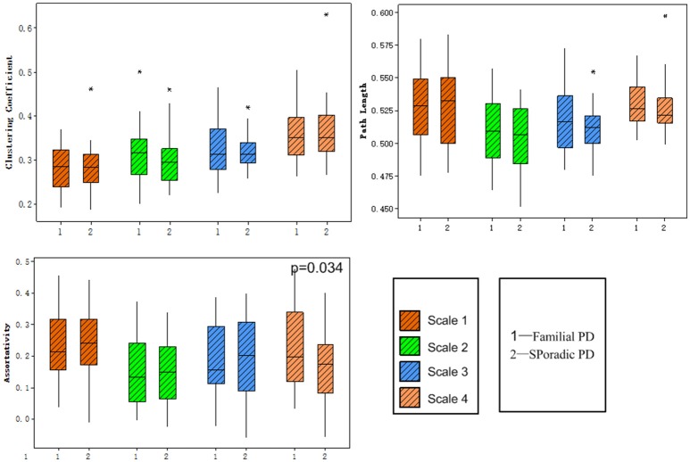
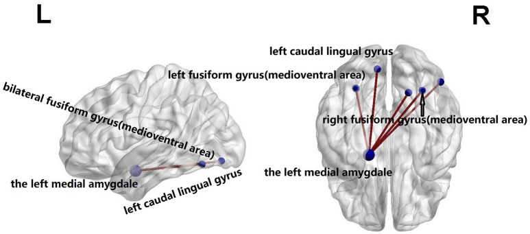
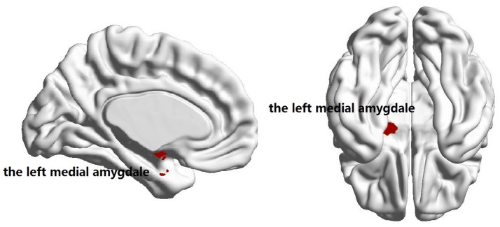
Familial Parkinson's disease (PD) is often caused by mutation of a certain gene, while sporadic PD is associated with variants of genes which can influence the susceptibility to PD. The goal of this study was to investigate the difference between the two forms of PD in terms of brain abnormalities using resting-state functional MRI and graph theory. Thirty-one familial PD patients and 36 sporadic PD patients underwent resting-state functional MRI scanning. Frequency-dependent functional connectivity was calculated for each subject using wavelet-based correlations of BOLD signal over 246 brain regions from Brainnetome Atlas. Graph theoretical analysis was then performed to analyze the topology of the functional network, and functional connectome differences were identified with a network-based statistical approach. Our results revealed a frequency-specific (0.016 and 0.031 Hz) connectome difference between familial and sporadic forms of PD, as indicated by an increase in assortativity and decrease in the nodal strength in the left medial amygdala of the familial PD group. In addition, the familial PD patients also showed a distinctive functional network between the left medial amygdala and regions related to retrieval of motion information. The present study indicates that the medial amygdala might be most vulnerable to both sporadic and familial PD. Our findings provide some new insights into disrupted resting-state functional connectomes between sporadic PD and familial PD.
家族性帕金森病(PD)通常由某一特定基因突变引起,而散发性PD则与可影响PD易感性的基因变异有关。本研究的目的是利用静息态功能磁共振成像(MRI)和图论,研究这两种形式的PD在脑异常方面的差异。31名家族性PD患者和36名散发性PD患者接受了静息态功能MRI扫描。使用基于小波的来自脑网络组图谱246个脑区的BOLD信号相关性,为每个受试者计算频率依赖性功能连接。然后进行图论分析以分析功能网络的拓扑结构,并采用基于网络的统计方法识别功能连接组差异。我们的结果显示,家族性和散发性PD之间存在频率特异性(0.016和0.031Hz)的连接组差异,家族性PD组左侧内侧杏仁核的聚类系数增加和节点强度降低表明了这一点。此外,家族性PD患者在左侧内侧杏仁核与与运动信息检索相关的区域之间还表现出独特的功能网络。本研究表明,内侧杏仁核可能对散发性和家族性PD都最为敏感。我们的发现为散发性PD和家族性PD之间静息态功能连接组的破坏提供了一些新的见解。