Department of Urology, University Hospital Carl Gustav Carus, Technische Universität Dresden, Fetscherstrasse 74, 01307, Dresden, Germany.
Department Radiopharmaceutical and Chemical Biology, Helmholtz-Zentrum Dresden-Rossendorf (HZDR), Institute of Radiopharmaceutical Cancer Research, Bautzner Landstrasse 400, 01328, Dresden, Germany.
BMC Cancer. 2017 Nov 23;17(1):790. doi: 10.1186/s12885-017-3778-3.
Novel theranostic options for high-risk non-muscle invasive bladder cancer are urgently needed. This requires a thorough evaluation of experimental approaches in animal models best possibly reflecting human disease before entering clinical studies. Although several bladder cancer xenograft models were used in the literature, the establishment of an orthotopic bladder cancer model in mice remains challenging.
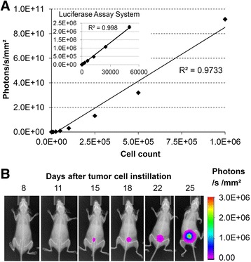
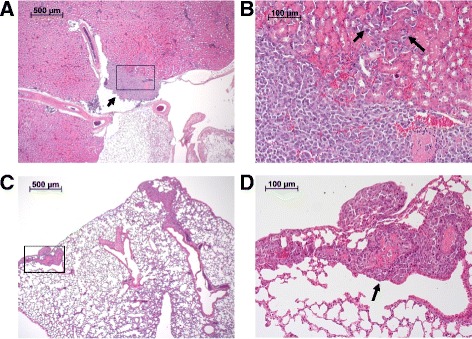
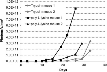
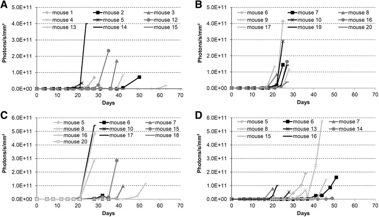
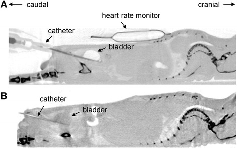
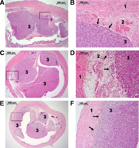
Luciferase-transduced UM-UC-3K1 bladder cancer cells were instilled transurethrally via 24G permanent venous catheters into athymic NMRI and BALB/c nude mice as well as into SCID-beige mice. Besides the mouse strain, the pretreatment of the bladder wall (trypsin or poly-L-lysine), tumor cell count (0.5 × 10-5.0 × 10) and tumor cell dwell time in the murine bladder (30 min - 2 h) were varied. Tumors were morphologically and functionally visualized using bioluminescence imaging (BLI), magnetic resonance imaging (MRI), and positron emission tomography (PET).
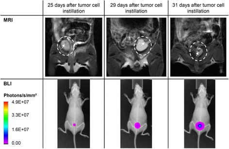
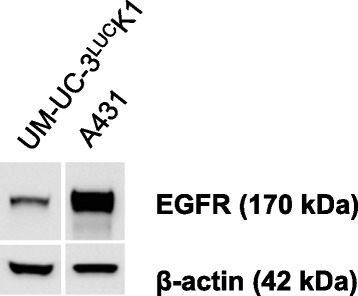
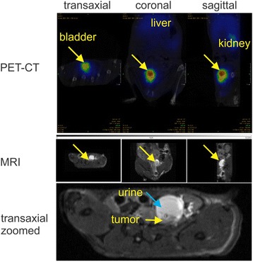
Immunodeficiency of the mouse strains was the most important factor influencing cancer cell engraftment, whereas modifying cell count and instillation time allowed fine-tuning of the BLI signal start and duration - both representing the possible treatment period for the evaluation of new therapeutics. Best orthotopic tumor growth was achieved by transurethral instillation of 1.0 × 10 UM-UC-3K1 bladder cancer cells into SCID-beige mice for 2 h after bladder pretreatment with poly-L-lysine. A pilot PET experiment using Ga-cetuximab as transurethrally administered radiotracer revealed functional expression of epidermal growth factor receptor as representative molecular characteristic of engrafted cancer cells in the bladder.
With the optimized protocol in SCID-beige mice an applicable and reliable model of high-risk non-muscle invasive bladder cancer for the development of novel theranostic approaches was established.
高危非肌肉浸润性膀胱癌迫切需要新的治疗方法。这需要在进入临床研究之前,在尽可能反映人类疾病的动物模型中彻底评估实验方法。尽管文献中已经使用了几种膀胱癌异种移植模型,但在小鼠中建立原位膀胱癌模型仍然具有挑战性。
通过 24G 永久静脉导管将荧光素酶转导的 UM-UC-3K1 膀胱癌细胞经尿道注入无胸腺 NMRI 和 BALB/c 裸鼠以及 SCID-beige 小鼠。除了小鼠品系外,还改变了膀胱壁预处理(胰蛋白酶或多聚赖氨酸)、肿瘤细胞计数(0.5×10-5.0×10)和肿瘤细胞在小鼠膀胱中的停留时间(30 分钟至 2 小时)。使用生物发光成像(BLI)、磁共振成像(MRI)和正电子发射断层扫描(PET)对肿瘤进行形态和功能可视化。
小鼠品系的免疫缺陷是影响癌细胞植入的最重要因素,而改变细胞计数和注入时间可以微调 BLI 信号的起始和持续时间——这两个因素都代表了评估新疗法的可能治疗期。通过经尿道注入 1.0×10 个 UM-UC-3K1 膀胱癌细胞,并用多聚赖氨酸预处理膀胱 2 小时,在 SCID-beige 小鼠中实现了最佳的原位肿瘤生长。使用 Ga-西妥昔单抗作为经尿道给予的放射性示踪剂进行的初步 PET 实验显示,表皮生长因子受体的功能性表达是膀胱中植入癌细胞的代表性分子特征。
通过在 SCID-beige 小鼠中优化方案,建立了一种适用于高危非肌肉浸润性膀胱癌的新型治疗方法的可行且可靠的模型。