• 文献检索
  • 文档翻译
  • 深度研究
  • 学术资讯
  • Suppr Zotero 插件Zotero 插件
  • 邀请有礼
  • 套餐&价格
  • 历史记录
应用&插件
Suppr Zotero 插件Zotero 插件浏览器插件Mac 客户端Windows 客户端微信小程序
定价
高级版会员购买积分包购买API积分包
服务
文献检索文档翻译深度研究API 文档MCP 服务
关于我们
关于 Suppr公司介绍联系我们用户协议隐私条款
关注我们

Suppr 超能文献

核心技术专利:CN118964589B侵权必究
粤ICP备2023148730 号-1Suppr @ 2026

文献检索

告别复杂PubMed语法,用中文像聊天一样搜索,搜遍4000万医学文献。AI智能推荐,让科研检索更轻松。

立即免费搜索

文件翻译

保留排版,准确专业,支持PDF/Word/PPT等文件格式,支持 12+语言互译。

免费翻译文档

深度研究

AI帮你快速写综述,25分钟生成高质量综述,智能提取关键信息,辅助科研写作。

立即免费体验

比较微计算机断层扫描和组织形态计量学评价聚醚醚酮涂层钛植入物在大鼠模型中的骨整合。

A comparison of micro-CT and histomorphometry for evaluation of osseointegration of PEO-coated titanium implants in a rat model.

机构信息

Department of Orthopedics, Ren Ji Hospital, School of Medicine, Shanghai Jiao Tong University, Shanghai, 200127, P.R. China.

State Key Laboratory of High Performance Ceramics and Superfine Microstructure, Shanghai Institute of Ceramics, Chinese Academy of Sciences, Shanghai, 200050, P.R. China.

出版信息

Sci Rep. 2017 Nov 24;7(1):16270. doi: 10.1038/s41598-017-16465-4.

DOI:10.1038/s41598-017-16465-4
PMID:29176604
原文链接:https://pmc.ncbi.nlm.nih.gov/articles/PMC5701240/
Abstract

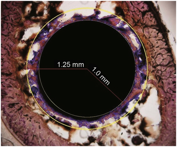
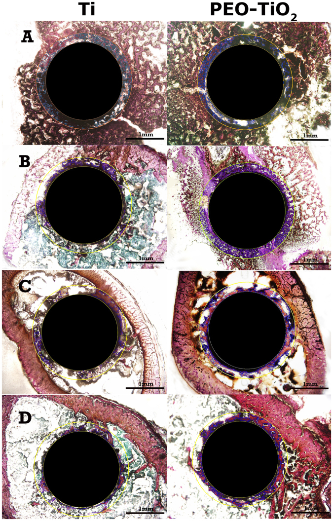
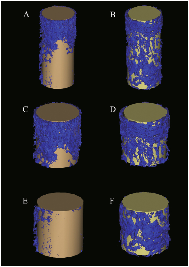
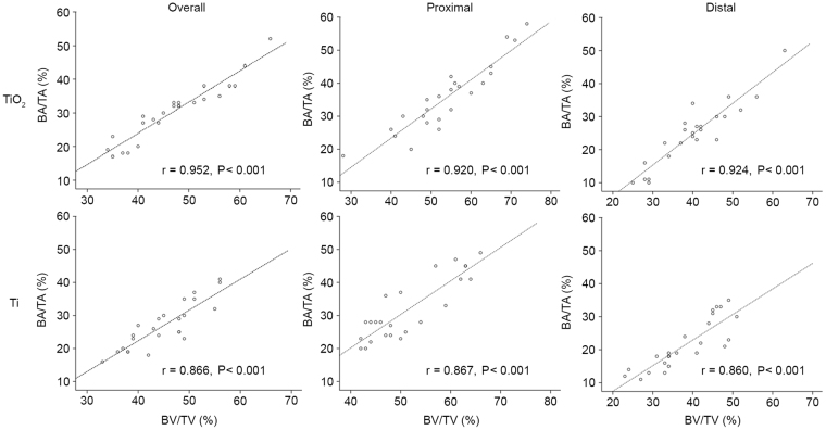
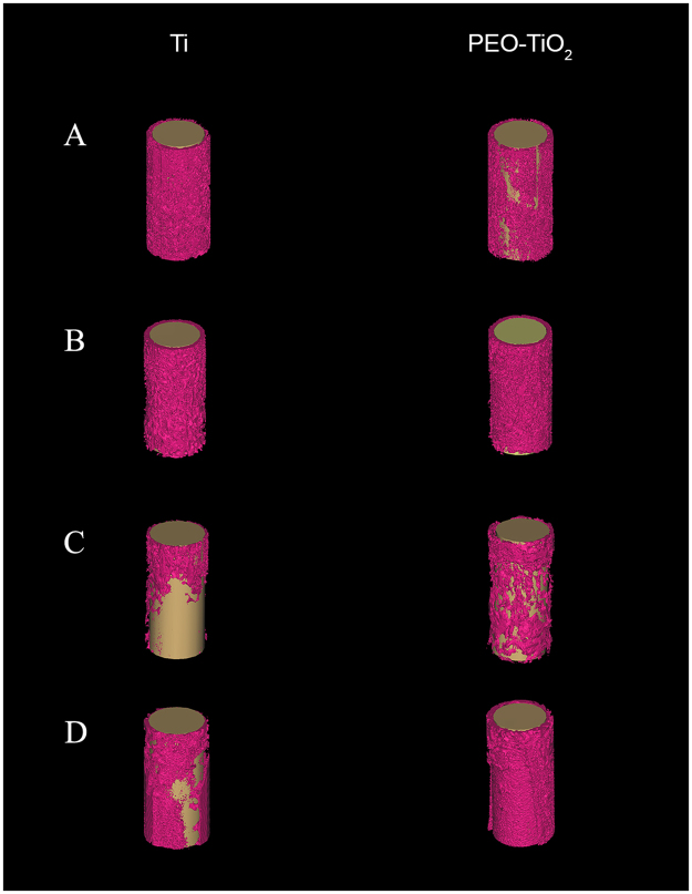
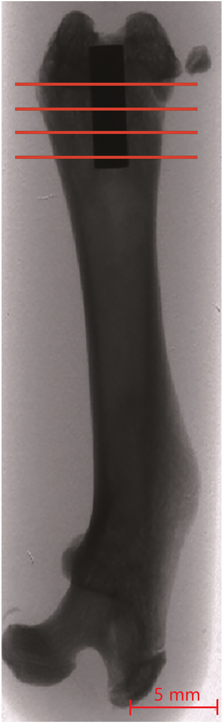
The aim of the present study was to determine the correlation between bone volume density (BV/TV) around a titanium implant determined by micro-computed tomography (micro-CT) and bone area density (BA/TA) measurements obtained using histomorphometry. An intramedullary rat femur implant model was evaluated to compare raw titanium implants with plasma electrolytic oxidation (PEO)-coated titanium implants. Titanium and PEO-treated titanium pins were inserted into rat femurs under general anesthesia. The animals were sacrificed and femurs harvested at 0, 2, 4 and 6 weeks, and subsequently, histomorphometry and micro-CT were performed. BV/TV and BA/TA values were strongly and positively correlated at all time points and locations (with all correlation coefficients being >0.8 and with P < 0.001). BV/TV and BA/TA were significantly higher proximal to the growth plate than distal to the growth plate, with estimated differences of 14.10% (P < 0.001) and 11.95% (P < 0.001), respectively. BV/TV and BA/TA were significantly higher on the PEO-coated surface than on the raw titanium surface, with estimated differences of 3.20% (P = 0.044) and 4.10% (P = 0.018), respectively. Therefore, quantitative micro-CT analysis of BV/TV is correlated with BA/TA determined by histomorphometry when artifacts around titanium implants are minimized by a region of interest modification.

摘要

本研究旨在确定微计算机断层扫描(micro-CT)测定的钛种植体周围骨体积密度(BV/TV)与组织形态计量学获得的骨面积密度(BA/TA)测量值之间的相关性。评估了一种髓内大鼠股骨植入模型,以比较原始钛植入物和等离子电解氧化(PEO)涂层钛植入物。在全身麻醉下将钛和 PEO 处理的钛针插入大鼠股骨。在 0、2、4 和 6 周时处死动物并收获股骨,随后进行组织形态计量学和 micro-CT 检查。在所有时间点和位置,BV/TV 和 BA/TA 值均呈强正相关(所有相关系数均>0.8,P<0.001)。与生长板远端相比,生长板近端的 BV/TV 和 BA/TA 值显著更高,估计差异分别为 14.10%(P<0.001)和 11.95%(P<0.001)。PEO 涂层表面的 BV/TV 和 BA/TA 值明显高于原始钛表面,估计差异分别为 3.20%(P=0.044)和 4.10%(P=0.018)。因此,当通过感兴趣区域修改最小化钛植入物周围的伪影时,BV/TV 的定量 micro-CT 分析与组织形态计量学确定的 BA/TA 相关。

https://cdn.ncbi.nlm.nih.gov/pmc/blobs/a7aa/5701240/a8a09c11d962/41598_2017_16465_Fig7_HTML.jpg
https://cdn.ncbi.nlm.nih.gov/pmc/blobs/a7aa/5701240/f39463beab35/41598_2017_16465_Fig1_HTML.jpg
https://cdn.ncbi.nlm.nih.gov/pmc/blobs/a7aa/5701240/f7446f42a245/41598_2017_16465_Fig2_HTML.jpg
https://cdn.ncbi.nlm.nih.gov/pmc/blobs/a7aa/5701240/2ac05d677602/41598_2017_16465_Fig3_HTML.jpg
https://cdn.ncbi.nlm.nih.gov/pmc/blobs/a7aa/5701240/2cbc5d55db7d/41598_2017_16465_Fig4_HTML.jpg
https://cdn.ncbi.nlm.nih.gov/pmc/blobs/a7aa/5701240/835c77a72e09/41598_2017_16465_Fig5_HTML.jpg
https://cdn.ncbi.nlm.nih.gov/pmc/blobs/a7aa/5701240/67d0a425ef2c/41598_2017_16465_Fig6_HTML.jpg
https://cdn.ncbi.nlm.nih.gov/pmc/blobs/a7aa/5701240/a8a09c11d962/41598_2017_16465_Fig7_HTML.jpg

相似文献

1
A comparison of micro-CT and histomorphometry for evaluation of osseointegration of PEO-coated titanium implants in a rat model.比较微计算机断层扫描和组织形态计量学评价聚醚醚酮涂层钛植入物在大鼠模型中的骨整合。
Sci Rep. 2017 Nov 24;7(1):16270. doi: 10.1038/s41598-017-16465-4.
2
Enhancing osseointegration of titanium implants through large-grit sandblasting combined with micro-arc oxidation surface modification.通过大颗粒喷砂联合微弧氧化表面改性增强钛植入物的骨整合。
J Mater Sci Mater Med. 2019 Jun 11;30(6):73. doi: 10.1007/s10856-019-6276-0.
3
Comparison of the effects of phosphate-coated and sandblasted acid-etched titanium implants on osseointegration: a microcomputed tomographic examination in the canine model.磷酸钙涂层和喷砂酸蚀钛种植体对骨整合影响的比较:犬模型中的微计算机断层扫描检查。
Int J Oral Maxillofac Implants. 2012 Sep-Oct;27(5):1069-80.
4
[The acceleration of titanium implant osseointegration by low intensity pulsed ultrasound: an experimental study in rats].[低强度脉冲超声促进钛种植体骨结合的实验研究:大鼠实验]
Zhonghua Kou Qiang Yi Xue Za Zhi. 2011 Jul;46(7):425-30.
5
Effects of cisplatin chemotherapy on the osseointegration of titanium implants.顺铂化疗对钛植入物骨整合的影响。
J Craniomaxillofac Surg. 2016 Apr;44(4):337-46. doi: 10.1016/j.jcms.2016.01.012. Epub 2016 Jan 22.
6
The effect of systemic nicotine on osseointegration of titanium implants in the rat femur.全身尼古丁对大鼠股骨钛植入物骨整合的影响。
J Oral Implantol. 2010;36(3):185-93. doi: 10.1563/AAID-JOI-D-09-00050.
7
A technique for evaluating bone ingrowth into 3D printed, porous Ti6Al4V implants accurately using X-ray micro-computed tomography and histomorphometry.一种利用X射线显微计算机断层扫描和组织形态计量学精确评估骨长入3D打印多孔Ti6Al4V植入物的技术。
Micron. 2017 Mar;94:1-8. doi: 10.1016/j.micron.2016.11.009. Epub 2016 Nov 21.
8
The reliability of cone-beam computed tomography to assess bone density at dental implant recipient sites: a histomorphometric analysis by micro-CT.锥形束计算机断层扫描评估种植体接受部位骨密度的可靠性:微 CT 组织形态计量学分析。
Clin Oral Implants Res. 2013 Aug;24(8):871-9. doi: 10.1111/j.1600-0501.2011.02390.x. Epub 2012 Jan 17.
9
Effect of lycopene on titanium implant osseointegration in ovariectomized rats.番茄红素对去卵巢大鼠钛种植体骨整合的影响。
J Orthop Surg Res. 2018 Sep 17;13(1):237. doi: 10.1186/s13018-018-0944-5.
10
Parathyroid hormone 1-34 enhances titanium implant anchorage in low-density trabecular bone: a correlative micro-computed tomographic and biomechanical analysis.甲状旁腺激素1-34增强钛植入物在低密度小梁骨中的锚固:一项相关的微观计算机断层扫描和生物力学分析。
Bone. 2006 Aug;39(2):276-82. doi: 10.1016/j.bone.2006.02.004. Epub 2006 Apr 17.

引用本文的文献

1
Recapitulation of angiogenesis and osteogenesis within an muscle pouch-based coral-derived macroporous construct organoid model.基于肌肉袋的珊瑚衍生大孔构建体类器官模型内血管生成和成骨作用的概述
J Orthop Translat. 2025 Apr 26;52:478-491. doi: 10.1016/j.jot.2025.04.002. eCollection 2025 May.
2
mRNA encoding bone morphogenetic protein-2 facilitated peri-implant bone formation of titanium implants placed in rat femurs.编码骨形态发生蛋白-2的信使核糖核酸促进了置于大鼠股骨中的钛植入物周围的骨形成。
Sci Rep. 2025 May 22;15(1):17852. doi: 10.1038/s41598-025-02931-x.
3
Effect of Ti6Al4V Alloy Surface and Porosity on Bone Osseointegration: In Vivo Pilot Study in Rabbits.

本文引用的文献

1
Biomechanical, micro-computed tomographic and immunohistochemical analysis of early osseous integration at titanium implants placed following lateral ridge augmentation using extracted tooth roots.使用拔牙牙根进行侧方牙槽嵴增高术后植入钛种植体早期骨结合的生物力学、显微计算机断层扫描及免疫组织化学分析
Clin Oral Implants Res. 2017 Mar;28(3):334-340. doi: 10.1111/clr.12803. Epub 2016 Mar 29.
2
Intermittent Administration of Parathyroid Hormone [1-34] Prevents Particle-Induced Periprosthetic Osteolysis in a Rat Model.甲状旁腺激素[1-34]间歇性给药可预防大鼠模型中颗粒诱导的假体周围骨溶解。
PLoS One. 2015 Oct 6;10(10):e0139793. doi: 10.1371/journal.pone.0139793. eCollection 2015.
3
Ti6Al4V合金表面及孔隙率对骨整合的影响:家兔体内初步研究
Materials (Basel). 2025 May 6;18(9):2141. doi: 10.3390/ma18092141.
4
Evaluation of osseointegration of plasma treated polyaryletherketone maxillofacial implants.等离子体处理的聚芳醚酮颌面植入物的骨整合评估。
Sci Rep. 2025 Jan 13;15(1):1895. doi: 10.1038/s41598-024-80335-z.
5
A two-step hydrothermal method for micro/nanotextured titanium implants and their integration outcomes in goat mandible.一种用于制备微/纳米纹理化钛植入物及其在山羊下颌骨中整合效果的两步水热法。
Nanomedicine (Lond). 2025 Jan;20(1):53-62. doi: 10.1080/17435889.2024.2435246. Epub 2024 Dec 2.
6
Analysis to evaluate novel separable dental implant stability: An experimental study in rabbits.评估新型可分离式牙种植体稳定性的分析:一项在兔子身上的实验研究。
J Dent Sci. 2024 Oct;19(4):2286-2294. doi: 10.1016/j.jds.2024.02.018. Epub 2024 Feb 26.
7
Radiographic texture of the trabecular bone of the proximal phalanx in horses with metacarpophalangeal osteoarthritis.掌指关节骨关节炎马匹近端指骨小梁骨的放射学纹理
J Equine Sci. 2024 Jul;35(2):21-28. doi: 10.1294/jes.35.21. Epub 2024 Jul 3.
8
Magnesium promotes vascularization and osseointegration in diabetic states.镁促进糖尿病状态下的血管生成和骨整合。
Int J Oral Sci. 2024 Jan 31;16(1):10. doi: 10.1038/s41368-023-00271-y.
9
Assessing bone formation on hydrophilic and hydrophobic implant surfaces in a murine model treated with bisphosphonates.评估在应用双膦酸盐治疗的小鼠模型中亲水和疏水种植体表面的成骨情况。
Clin Oral Investig. 2024 Jan 13;28(1):89. doi: 10.1007/s00784-023-05480-7.
10
Comparative evaluation of 3D-printed and conventional implants in vivo: a quantitative microcomputed tomographic and histomorphometric analysis.3D 打印与传统植入物体内对比评估:定量微计算机断层扫描和组织形态计量学分析。
Sci Rep. 2023 Nov 29;13(1):21041. doi: 10.1038/s41598-023-48315-x.
A special healing pattern in stable metaphyseal fractures.
稳定型干骺端骨折的一种特殊愈合模式。
Acta Orthop. 2015 Apr;86(2):238-42. doi: 10.3109/17453674.2014.1003127. Epub 2015 Jan 13.
4
Computational analysis of primary implant stability in trabecular bone.松质骨中种植体初期稳定性的计算分析
J Biomech. 2015 Mar 18;48(5):807-15. doi: 10.1016/j.jbiomech.2014.12.008. Epub 2014 Dec 29.
5
Evaluation of Osseointegration around Tibial Implants in Rats by Ibandronate-Treated Nanotubular Ti-32Nb-5Zr Alloy.依班膦酸处理的纳米管Ti-32Nb-5Zr合金对大鼠胫骨植入物周围骨整合的评估
Biomol Ther (Seoul). 2014 Nov;22(6):563-9. doi: 10.4062/biomolther.2014.111. Epub 2014 Nov 30.
6
Microcomputed tomographic and biomechanical analysis of orthodontic microscrew stability under continuous or intermittent force.连续或间歇力作用下正畸微螺钉稳定性的显微计算机断层扫描及生物力学分析
Int J Oral Maxillofac Implants. 2014 Jul-Aug;29(4):819-25. doi: 10.11607/jomi.3191.
7
Metal release from ceramic coatings for dental implants.牙科植入物陶瓷涂层的金属释放。
Dent Mater. 2014 Mar;30(3):e28-40. doi: 10.1016/j.dental.2013.12.011. Epub 2014 Jan 14.
8
Utilizing micro-computed tomography to evaluate bone structure surrounding dental implants: a comparison with histomorphometry.利用微计算机断层扫描评估种植牙周围的骨结构:与组织形态计量学的比较。
J Biomed Mater Res B Appl Biomater. 2013 Oct;101(7):1259-66. doi: 10.1002/jbm.b.32938. Epub 2013 May 10.
9
Histological and synchrotron radiation-based computed microtomography study of 2 human-retrieved direct laser metal formed titanium implants.对 2 个人体取出的直接激光金属成型钛植入物进行组织学和同步辐射计算机微断层扫描研究。
Implant Dent. 2013 Apr;22(2):175-81. doi: 10.1097/ID.0b013e318282817d..
10
Mathematical filtering minimizes metallic halation of titanium implants in MicroCT images.数学滤波可将微型计算机断层扫描(MicroCT)图像中钛植入物的金属光晕降至最低。
J Calif Dent Assoc. 2013 Jan;41(1):41-5.