Reiner D S, Douglas H, Gillin F D
Department of Pathology, University of California, San Diego 92103.
Infect Immun. 1989 Mar;57(3):963-8. doi: 10.1128/iai.57.3.963-968.1989.
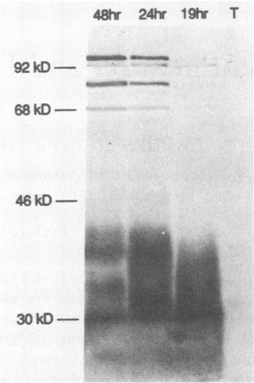
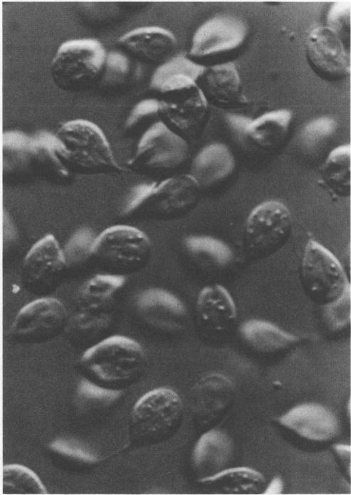
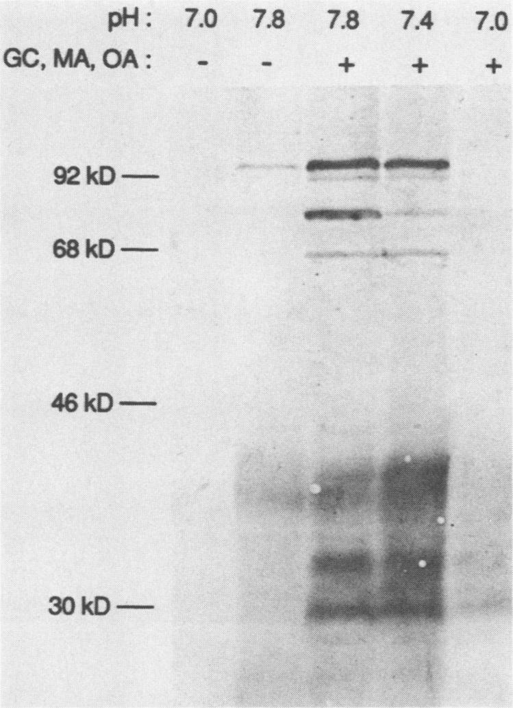
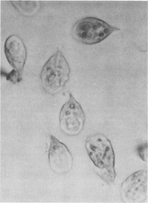
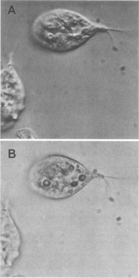
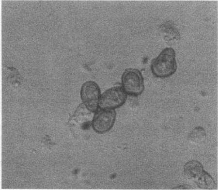
We induced Giardia lamblia trophozoites to encyst in vitro by exposure to conditions which are specific to the human small intestinal milieu. We now show that encystation entails the appearance of two new groups of antigens detected in Western blots by rabbit antiserum against cysts which had been purified from human feces. A heterodisperse group of lower-molecular-mass antigens (approximately 21 to 39 kilodaltons) was expressed relatively early (less than 19 h) in encystation. In contrast, discrete bands at approximately 66, 78, 92, and 103 kilodaltons only appeared after 24 h of incubation under conditions which lead to production of large numbers of water-resistant cysts. We also describe for the first time the appearance of prominent cytoplasmic vesicles, which were the earliest morphologic change in encysting trophozoites observable by light microscopy. Early in encystation, cyst wall antigens were concentrated in these vesicles, as shown by immunocytochemistry, suggesting that the vesicles function in export of cyst wall constituents.
我们通过将蓝氏贾第鞭毛虫滋养体暴露于模拟人类小肠环境的特定条件下,诱导其在体外形成包囊。我们现在发现,包囊化过程会出现两组新的抗原,这些抗原可通过兔抗人粪便纯化包囊血清在蛋白质免疫印迹法中检测到。一组低分子量的异质抗原(约21至39千道尔顿)在包囊化过程中相对较早(少于19小时)表达。相比之下,只有在导致大量耐水包囊产生的条件下孵育24小时后,约66、78、92和103千道尔顿的离散条带才会出现。我们还首次描述了显著的细胞质囊泡的出现,这是光镜下可观察到的包囊化滋养体最早的形态学变化。在包囊化早期,免疫细胞化学显示包囊壁抗原集中在这些囊泡中,这表明这些囊泡参与包囊壁成分的输出。