Department of Nephrology and Hypertension, Glickman Urological and Kidney Institute, Cleveland Clinic, Cleveland, Ohio.
Department of Pathobiology, Lerner Research Institute, Cleveland Clinic, Cleveland, Ohio.
J Am Soc Nephrol. 2018 Mar;29(3):869-879. doi: 10.1681/ASN.2016121322. Epub 2017 Nov 27.
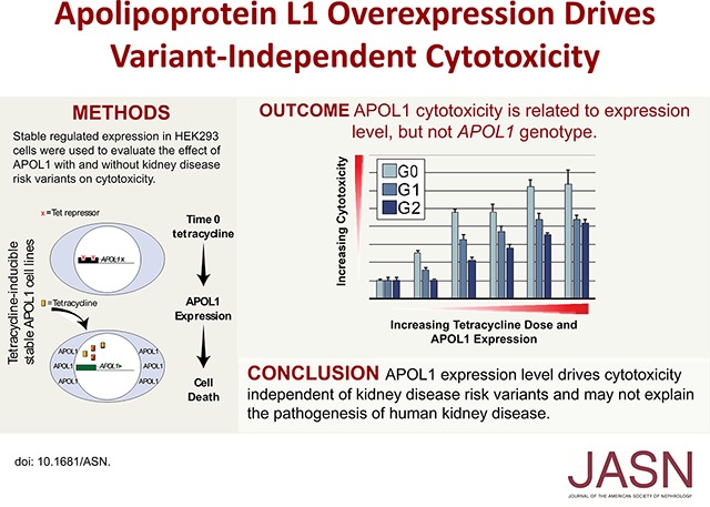
Coding variants in the gene are associated with kidney diseases in African ancestral populations; yet, the underlying biologic mechanisms remain uncertain. Variant-dependent autophagic and cytotoxic cell death have been proposed as pathogenic pathways mediating kidney injury. To examine this possibility, we conditionally expressed APOL1-G0 (reference), -G1, and -G2 (variants) using a tetracycline-regulated system in HEK293 cells. Autophagy was monitored biochemically and cell death was measured using multiple assays. We measured intracellular Na and K content with atomic absorption spectroscopy and APOL1-dependent currents with whole-cell patch clamping. Neither reference nor variant APOL1s induced autophagy. At high expression levels, APOL1-G0, -G1, and -G2 inserted into the plasma membrane and formed pH-sensitive cation channels, causing collapse of cellular Na and K gradients, phosphorylation of p38 mitogen-activated protein kinase, and cell death, without variant-dependent differences. APOL1-G0 and -G2 exhibited similar channel properties in whole-cell patch clamp experiments. At low expression levels, neither reference nor variant APOL1s localized on the plasma membrane, Na and K gradients were maintained, and cells remained viable. Our results indicate that APOL1-mediated pore formation is critical for the trypanolytic activity of APOL1 and drives APOL1-mediated cytotoxicity in overexpression systems. The absence of cytotoxicity at physiologic expression levels suggests variant-dependent intracellular K loss and cytotoxicity does not drive kidney disease progression.
基因中的编码变异与非洲裔人群的肾脏疾病有关;然而,潜在的生物学机制仍不确定。依赖于变异的自噬和细胞毒性细胞死亡已被提议作为介导肾脏损伤的致病途径。为了研究这种可能性,我们使用四环素调控系统在 HEK293 细胞中条件表达了 APOL1-G0(参照)、-G1 和 -G2(变异体)。通过生化方法监测自噬,并用多种测定方法测量细胞死亡。我们用原子吸收光谱法测量细胞内 Na 和 K 的含量,用全细胞膜片钳技术测量 APOL1 依赖性电流。参照和变异体 APOL1 均未诱导自噬。在高表达水平下,APOL1-G0、-G1 和 -G2 插入质膜并形成 pH 敏感的阳离子通道,导致细胞内 Na 和 K 梯度崩溃、p38 丝裂原激活蛋白激酶磷酸化和细胞死亡,而无变异依赖性差异。APOL1-G0 和 -G2 在全细胞膜片钳实验中表现出相似的通道特性。在低表达水平下,参照和变异体 APOL1 均未定位于质膜,Na 和 K 梯度得以维持,细胞仍然存活。我们的结果表明,APOL1 介导的孔形成对于 APOL1 的杀变形虫活性至关重要,并在过表达系统中驱动 APOL1 介导的细胞毒性。在生理表达水平下缺乏细胞毒性表明变异体依赖性细胞内 K 丢失,并且细胞毒性不会导致肾脏疾病进展。