Cui Shaohua, Su Xinying, Dong Lili, Qian Jialin, Ye Lin, Zhang Tianwei, Fu Haihua, Han Hulin, Huang Jiaqi, Yao Yihong, Gu Yi, Jiang Liyan
Department of Respiratory Medicine, Shanghai Chest Hospital, Shanghai Jiao Tong University, Shanghai, China.
Asia & Emerging Markets, iMed, AstraZeneca, Shanghai, China.
J Cancer. 2017 Oct 24;8(19):4075-4082. doi: 10.7150/jca.21415. eCollection 2017.
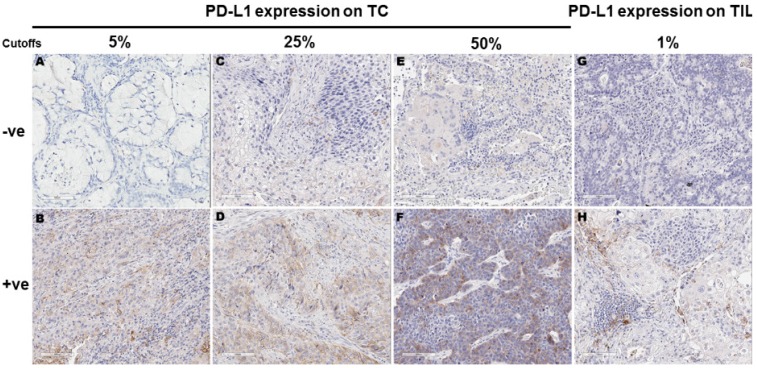
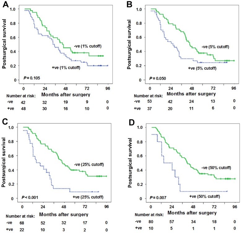
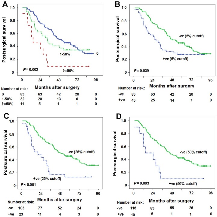
To investigate the relationship between programmed death ligand 1 (PD-L1) expression using 5%, 25%, 50% cutoffs in tumor cells (TC) and postsurgical survival in non-small-cell lung cancer (NSCLC) patients. For samples with tumor infiltrating lymphocytes (TIL), correlation between PD-L1 expression in TIL using 1% cutoff and postsurgical survival was also evaluated. Primary NSCLC tumor surgical samples staging I to IIIA of 126 patients who underwent surgical procedures from September 2009 to August 2012 in Shanghai Chest Hospital, Shanghai Jiao Tong University were retrospectively included. PD-L1 protein expression was detected by immunohistochemistry (IHC) assays. A rabbit anti-human PD-L1 (E1L3N) monoclonal antibody (1:300, CST#13684, Cell Signaling Technology) was used for PD-L1 IHC staining. PD-L1 expression was evaluated both on TC and TIL. Univariate and multivariate analyses for postsurgical survival were done using Kaplan-Meier and Cox regression model, respectively. The median postsurgical survival for all patients was 44.1 months [95% confidence interval (CI): 33.9-70.0 months). The median postsurgical survival for PD-L1 expression percentage 0, 1-50% and ≥50% were 51.9 months (95%CI: 33.9-70.0 months), 33.2 months (95%CI: 20.8-45.6 months) and 14.7 months (95%CI: 1.9-27.6 months), respectively ( = 0.002). Clinical stage and PD-L1 expression in TC (25% cutoff or 50% cutoff values) were found to be independent predictors for longer postsurgical survival in all cohort. Ninety (71.4%) of the 126 samples were identified to concurrent TIL. The median postsurgical survival time was 39.6 months (95% CI: 31.8-47.4 months) in patients with TIL. PD-L1 expression in TC (25% cutoff or 50% cutoff values) was found to be the independent predictor for longer postsurgical survival time in patients with TIL. PD-L1 negative expression in TC at 25% or 50% cutoff values was the independent predictor for longer postsurgical survival time in both NSCLC samples and NSCLC samples with TIL. For patients with PD-L1 high expression at 25% or 50% cutoff values, PD-L1 blocking may be considered.
探讨采用5%、25%、50%的截断值评估肿瘤细胞(TC)中程序性死亡配体1(PD-L1)表达与非小细胞肺癌(NSCLC)患者术后生存之间的关系。对于存在肿瘤浸润淋巴细胞(TIL)的样本,还评估了采用1%截断值评估的TIL中PD-L1表达与术后生存的相关性。回顾性纳入了2009年9月至2012年8月在上海交通大学附属上海胸科医院接受手术的126例I至IIIA期原发性NSCLC肿瘤手术样本。通过免疫组织化学(IHC)检测PD-L1蛋白表达。使用兔抗人PD-L1(E1L3N)单克隆抗体(1:300,CST#13684,Cell Signaling Technology)进行PD-L1 IHC染色。在TC和TIL上均评估PD-L1表达。分别采用Kaplan-Meier法和Cox回归模型对术后生存进行单因素和多因素分析。所有患者的术后生存中位数为44.1个月[95%置信区间(CI):33.9 - 70.0个月]。PD-L1表达百分比为0、1 - 50%和≥50%的患者术后生存中位数分别为51.9个月(95%CI:33.9 - 70.0个月)、33.2个月(95%CI:20.8 - 45.6个月)和14.7个月(95%CI:1.9 - 27.6个月)(P = 0.002)。临床分期和TC中PD-L1表达(25%或50%截断值)被发现是所有队列中术后生存时间更长的独立预测因素。126个样本中有90个(71.4%)同时存在TIL。存在TIL的患者术后生存时间中位数为39.6个月(95%CI:31.8 - 47.4个月)。TC中PD-L1表达(25%或50%截断值)被发现是存在TIL患者术后生存时间更长的独立预测因素。TC中25%或50%截断值时PD-L1阴性表达是NSCLC样本和存在TIL的NSCLC样本中术后生存时间更长的独立预测因素。对于25%或50%截断值时PD-L1高表达的患者,可考虑进行PD-L1阻断治疗。