• 文献检索
  • 文档翻译
  • 深度研究
  • 学术资讯
  • Suppr Zotero 插件Zotero 插件
  • 邀请有礼
  • 套餐&价格
  • 历史记录
应用&插件
Suppr Zotero 插件Zotero 插件浏览器插件Mac 客户端Windows 客户端微信小程序
定价
高级版会员购买积分包购买API积分包
服务
文献检索文档翻译深度研究API 文档MCP 服务
关于我们
关于 Suppr公司介绍联系我们用户协议隐私条款
关注我们

Suppr 超能文献

核心技术专利:CN118964589B侵权必究
粤ICP备2023148730 号-1Suppr @ 2026

文献检索

告别复杂PubMed语法,用中文像聊天一样搜索,搜遍4000万医学文献。AI智能推荐,让科研检索更轻松。

立即免费搜索

文件翻译

保留排版,准确专业,支持PDF/Word/PPT等文件格式,支持 12+语言互译。

免费翻译文档

深度研究

AI帮你快速写综述,25分钟生成高质量综述,智能提取关键信息,辅助科研写作。

立即免费体验

斑马鱼作为一种用于分析哺乳动物和天然晶状体蛋白启动子功能的模型系统。

The zebrafish as a model system for analyzing mammalian and native -crystallin promoter function.

作者信息

Posner Mason, Murray Kelly L, McDonald Matthew S, Eighinger Hayden, Andrew Brandon, Drossman Amy, Haley Zachary, Nussbaum Justin, David Larry L, Lampi Kirsten J

机构信息

Department of Biology/Toxicology, Ashland University, Ashland, OH, United States of America.

Department of Biology, Lakeland Community College, Kirtland, OH, United States of America.

出版信息

PeerJ. 2017 Nov 27;5:e4093. doi: 10.7717/peerj.4093. eCollection 2017.

DOI:10.7717/peerj.4093
PMID:29201567
原文链接:https://pmc.ncbi.nlm.nih.gov/articles/PMC5708185/
Abstract

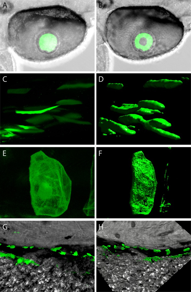
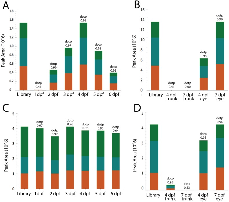
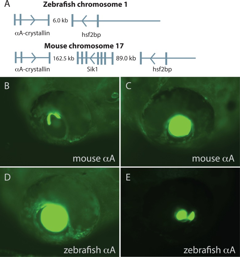
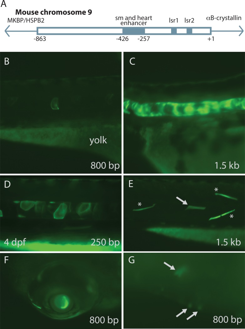
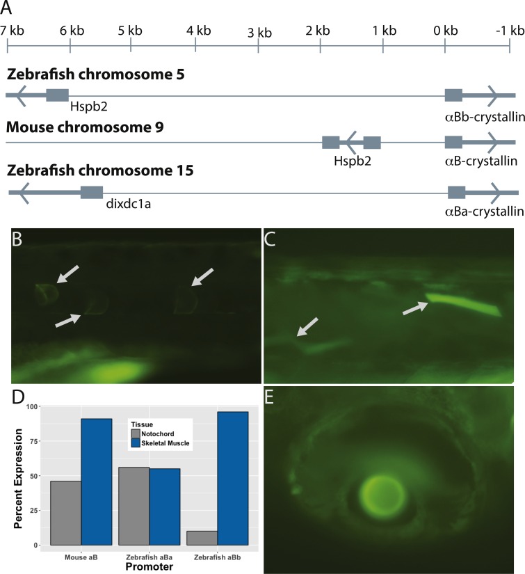
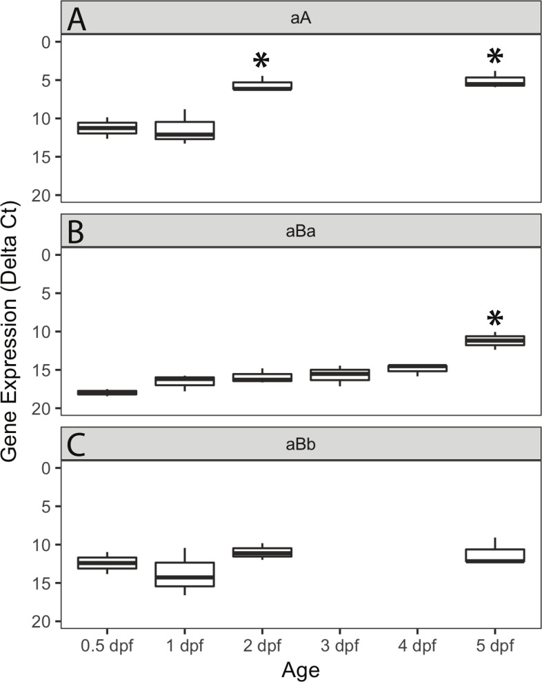
Previous studies have used the zebrafish to investigate the biology of lens crystallin proteins and their roles in development and disease. However, little is known about zebrafish α-crystallin promoter function, how it compares to that of mammals, or whether mammalian α-crystallin promoter activity can be assessed using zebrafish embryos. We injected a variety of α-crystallin promoter fragments from each species combined with the coding sequence for green fluorescent protein (GFP) into zebrafish zygotes to determine the resulting spatiotemporal expression patterns in the developing embryo. We also measured mRNA levels and protein abundance for all three zebrafish α-crystallins. Our data showed that mouse and zebrafish αA-crystallin promoters generated similar GFP expression in the lens, but with earlier onset when using mouse promoters. Expression was also found in notochord and skeletal muscle in a smaller percentage of embryos. Mouse αB-crystallin promoter fragments drove GFP expression primarily in zebrafish skeletal muscle, with less common expression in notochord, lens, heart and in extraocular regions of the eye. A short fragment containing only a lens-specific enhancer region increased lens and notochord GFP expression while decreasing muscle expression, suggesting that the influence of mouse promoter control regions carries over into zebrafish embryos. The two paralogous zebrafish αB-crystallin promoters produced subtly different expression profiles, with the aBa promoter driving expression equally in notochord and skeletal muscle while the αBb promoter resulted primarily in skeletal muscle expression. Messenger RNA for zebrafish αA increased between 1 and 2 days post fertilization (dpf), αBa increased between 4 and 5 dpf, but Bb remained at baseline levels through 5 dpf. Parallel reaction monitoring (PRM) mass spectrometry was used to detect αA, aBa, and αBb peptides in digests of zebrafish embryos. In whole embryos, αA-crystallin was first detected by 2 dpf, peaked in abundance by 4-5 dpf, and was localized to the eye. αBa was detected in whole embryo at nearly constant levels from 1-6 dpf, was also localized primarily to the eye and its abundance in extraocular tissues decreased from 4-7 dpf. In contrast, due to its low abundance, no αBb protein could be detected in whole embryo, or dissected eye and extraocular tissues. Our results show that mammalian α-crystallin promoters can be efficiently screened in zebrafish embryos and that their controlling regions are well conserved. An ontogenetic shift in zebrafish aBa-crystallin promoter activity provides an interesting system for examining the evolution and control of tissue specificity. Future studies that combine these promoter based approaches with the expanding ability to engineer the zebrafish genome via techniques such as CRISPR/Cas9 will allow the manipulation of protein expression to test hypotheses about lens crystallin function and its relation to lens biology and disease.

摘要

以往的研究利用斑马鱼来研究晶状体晶状体蛋白的生物学特性及其在发育和疾病中的作用。然而,关于斑马鱼α-晶状体蛋白启动子的功能、与哺乳动物启动子功能的比较,或者是否可以使用斑马鱼胚胎评估哺乳动物α-晶状体蛋白启动子活性,我们所知甚少。我们将来自每个物种的多种α-晶状体蛋白启动子片段与绿色荧光蛋白(GFP)的编码序列相结合,注射到斑马鱼受精卵中,以确定发育胚胎中产生的时空表达模式。我们还测量了所有三种斑马鱼α-晶状体蛋白的mRNA水平和蛋白质丰度。我们的数据表明,小鼠和斑马鱼的αA-晶状体蛋白启动子在晶状体中产生了相似的GFP表达,但使用小鼠启动子时表达开始得更早。在较小比例的胚胎中,在脊索和骨骼肌中也发现了表达。小鼠αB-晶状体蛋白启动子片段主要在斑马鱼骨骼肌中驱动GFP表达,在脊索、晶状体、心脏和眼外区域的表达较少见。一个仅包含晶状体特异性增强子区域的短片段增加了晶状体和脊索中的GFP表达,同时降低了肌肉中的表达,这表明小鼠启动子控制区域的影响在斑马鱼胚胎中也存在。两个同源的斑马鱼αB-晶状体蛋白启动子产生了略有不同的表达谱,αBa启动子在脊索和骨骼肌中驱动表达的程度相同,而αBb启动子主要导致骨骼肌表达。斑马鱼αA的信使RNA在受精后1至2天(dpf)之间增加,αBa在4至5 dpf之间增加,但Bb在5 dpf之前一直保持在基线水平。平行反应监测(PRM)质谱法用于检测斑马鱼胚胎消化物中的αA、αBa和αBb肽段。在整个胚胎中,αA-晶状体蛋白在2 dpf时首次被检测到,在4至5 dpf时丰度达到峰值,并定位于眼睛。αBa在1至6 dpf的整个胚胎中以几乎恒定的水平被检测到,也主要定位于眼睛,其在眼外组织中的丰度在4至7 dpf之间下降。相比之下,由于其丰度较低,在整个胚胎、解剖的眼睛和眼外组织中均未检测到αBb蛋白。我们的结果表明,哺乳动物α-晶状体蛋白启动子可以在斑马鱼胚胎中进行有效筛选,并且它们的控制区域具有良好的保守性。斑马鱼αBa-晶状体蛋白启动子活性的个体发育转变为研究组织特异性的进化和控制提供了一个有趣的系统。未来将这些基于启动子的方法与通过CRISPR/Cas9等技术对斑马鱼基因组进行工程改造的不断扩展的能力相结合的研究,将允许对蛋白质表达进行操纵,以检验关于晶状体晶状体蛋白功能及其与晶状体生物学和疾病关系的假设。

https://cdn.ncbi.nlm.nih.gov/pmc/blobs/c03f/5708185/610a0c5769f0/peerj-05-4093-g006.jpg
https://cdn.ncbi.nlm.nih.gov/pmc/blobs/c03f/5708185/1e9155e2b5d8/peerj-05-4093-g001.jpg
https://cdn.ncbi.nlm.nih.gov/pmc/blobs/c03f/5708185/0e2da6279eb6/peerj-05-4093-g002.jpg
https://cdn.ncbi.nlm.nih.gov/pmc/blobs/c03f/5708185/e87f1de54098/peerj-05-4093-g003.jpg
https://cdn.ncbi.nlm.nih.gov/pmc/blobs/c03f/5708185/7864efc3e317/peerj-05-4093-g004.jpg
https://cdn.ncbi.nlm.nih.gov/pmc/blobs/c03f/5708185/9d0a6d52263f/peerj-05-4093-g005.jpg
https://cdn.ncbi.nlm.nih.gov/pmc/blobs/c03f/5708185/610a0c5769f0/peerj-05-4093-g006.jpg

相似文献

1
The zebrafish as a model system for analyzing mammalian and native -crystallin promoter function.斑马鱼作为一种用于分析哺乳动物和天然晶状体蛋白启动子功能的模型系统。
PeerJ. 2017 Nov 27;5:e4093. doi: 10.7717/peerj.4093. eCollection 2017.
2
Loss of αBa-crystallin, but not αA-crystallin, increases age-related cataract in the zebrafish lens.αB 晶状体蛋白的缺失而非 αA 晶状体蛋白的缺失会导致斑马鱼晶状体的年龄相关性白内障。
Exp Eye Res. 2024 Jul;244:109918. doi: 10.1016/j.exer.2024.109918. Epub 2024 May 3.
3
Loss of αBa-crystallin, but not αA-crystallin, increases age-related cataract in the zebrafish lens.αB-晶状体蛋白而非αA-晶状体蛋白的缺失会增加斑马鱼晶状体中与年龄相关的白内障。
bioRxiv. 2024 Jan 3:2024.01.03.574085. doi: 10.1101/2024.01.03.574085.
4
A conserved role of αA-crystallin in the development of the zebrafish embryonic lens.αA-晶体蛋白在斑马鱼胚胎晶状体发育中的保守作用。
Exp Eye Res. 2015 Sep;138:104-13. doi: 10.1016/j.exer.2015.07.001. Epub 2015 Jul 4.
5
Why does the zebrafish cloche mutant develop lens cataract?斑马鱼钟形帽突变体为什么会发生晶状体白内障?
PLoS One. 2019 Mar 12;14(3):e0211399. doi: 10.1371/journal.pone.0211399. eCollection 2019.
6
Impact of α-crystallin protein loss on zebrafish lens development.α-晶体蛋白缺失对斑马鱼晶状体发育的影响。
Exp Eye Res. 2023 Feb;227:109358. doi: 10.1016/j.exer.2022.109358. Epub 2022 Dec 23.
7
Species-Specific Structural and Functional Divergence of α-Crystallins: Zebrafish αBa- and Rodent αA(ins)-Crystallin Encode Activated Chaperones.α-晶体蛋白的物种特异性结构与功能差异:斑马鱼αBa-晶体蛋白和啮齿动物αA(ins)-晶体蛋白编码活化伴侣蛋白。
Biochemistry. 2015 Sep 29;54(38):5949-58. doi: 10.1021/acs.biochem.5b00678. Epub 2015 Sep 17.
8
Chick delta1-crystallin enhancer influences mouse alphaA-crystallin promoter activity in transgenic mice.鸡δ1-晶体蛋白增强子影响转基因小鼠中鼠αA-晶体蛋白启动子的活性。
Invest Ophthalmol Vis Sci. 2004 Nov;45(11):4083-90. doi: 10.1167/iovs.03-1270.
9
Loss of the small heat shock protein αA-crystallin does not lead to detectable defects in early zebrafish lens development.αA-晶体小热休克蛋白缺失不会导致早期斑马鱼晶状体发育出现可检测的缺陷。
Exp Eye Res. 2013 Nov;116:227-33. doi: 10.1016/j.exer.2013.09.007. Epub 2013 Sep 25.
10
Insertion of a Pax6 consensus binding site into the alphaA-crystallin promoter acts as a lens epithelial cell enhancer in transgenic mice.将一个Pax6共有结合位点插入αA-晶体蛋白启动子中,在转基因小鼠中可作为晶状体上皮细胞增强子发挥作用。
Invest Ophthalmol Vis Sci. 2004 Jun;45(6):1930-9. doi: 10.1167/iovs.03-0856.

引用本文的文献

1
Data-Independent Acquisition Parallel Accumulation-Serial Fragmentation (diaPASEF) Analysis of the Separated Zebrafish Lens Improves Identifications.斑马鱼晶状体分离后的数据非依赖采集平行累积-序列碎裂(diaPASEF)分析可提高鉴定效率。
J Am Soc Mass Spectrom. 2025 Jun 9. doi: 10.1021/jasms.5c00087.
2
Data-independent acquisition parallel accumulation-serial fragmentation (diaPASEF) analysis of the separated zebrafish lens improves identifications.对分离的斑马鱼晶状体进行数据非依赖型采集平行累积-序列碎裂(diaPASEF)分析可提高鉴定效率。
bioRxiv. 2025 Mar 15:2025.03.13.642445. doi: 10.1101/2025.03.13.642445.
3
The generation and characterization of a transgenic zebrafish line with lens-specific Cre expression.

本文引用的文献

1
Evolutionary Origins of Pax6 Control of Crystallin Genes.Pax6 调控晶体蛋白基因的进化起源。
Genome Biol Evol. 2017 Aug 1;9(8):2075-2092. doi: 10.1093/gbe/evx153.
2
Cloche is a bHLH-PAS transcription factor that drives haemato-vascular specification.克洛什(Cloche)是一个 bHLH-PAS 转录因子,它驱动造血血管的特化。
Nature. 2016 Jul 14;535(7611):294-8. doi: 10.1038/nature18614.
3
Parallel reaction monitoring using quadrupole-Orbitrap mass spectrometer: Principle and applications.使用四极杆-轨道阱质谱仪的平行反应监测:原理与应用
具有晶状体特异性 Cre 表达的转基因斑马鱼系的产生和特性。
Mol Vis. 2024 Mar 19;30:123-136. eCollection 2024.
4
Rapid and precise genome engineering in a naturally short-lived vertebrate.在一种自然寿命较短的脊椎动物中进行快速而精确的基因组工程。
Elife. 2023 May 9;12:e80639. doi: 10.7554/eLife.80639.
5
Impact of α-crystallin protein loss on zebrafish lens development.α-晶体蛋白缺失对斑马鱼晶状体发育的影响。
Exp Eye Res. 2023 Feb;227:109358. doi: 10.1016/j.exer.2022.109358. Epub 2022 Dec 23.
6
Single cell transcriptomics of the developing zebrafish lens and identification of putative controllers of lens development.斑马鱼晶状体发育的单细胞转录组学及潜在晶状体发育调控因子的鉴定。
Exp Eye Res. 2021 May;206:108535. doi: 10.1016/j.exer.2021.108535. Epub 2021 Mar 9.
7
α-Crystallins in the Vertebrate Eye Lens: Complex Oligomers and Molecular Chaperones.脊椎动物眼晶状体中的α-晶体蛋白:复杂的寡聚物和分子伴侣。
Annu Rev Phys Chem. 2021 Apr 20;72:143-163. doi: 10.1146/annurev-physchem-090419-121428. Epub 2020 Dec 15.
8
Tnni1b-ECR183-d2, an 87 bp cardiac enhancer of zebrafish.Tnni1b - ECR183 - d2,一种斑马鱼的87碱基对心脏增强子。
PeerJ. 2020 Nov 4;8:e10289. doi: 10.7717/peerj.10289. eCollection 2020.
9
Quantifying protein dynamics and stability in a living organism.定量研究活生物体中的蛋白质动态和稳定性。
Nat Commun. 2019 Mar 12;10(1):1179. doi: 10.1038/s41467-019-09088-y.
10
Why does the zebrafish cloche mutant develop lens cataract?斑马鱼钟形帽突变体为什么会发生晶状体白内障?
PLoS One. 2019 Mar 12;14(3):e0211399. doi: 10.1371/journal.pone.0211399. eCollection 2019.
Proteomics. 2016 Aug;16(15-16):2146-59. doi: 10.1002/pmic.201500543. Epub 2016 May 27.
4
Regulation of c-Maf and αA-Crystallin in Ocular Lens by Fibroblast Growth Factor Signaling.成纤维细胞生长因子信号通路对晶状体中c-Maf和αA-晶状体蛋白的调控
J Biol Chem. 2016 Feb 19;291(8):3947-58. doi: 10.1074/jbc.M115.705103. Epub 2015 Dec 30.
5
Species-Specific Structural and Functional Divergence of α-Crystallins: Zebrafish αBa- and Rodent αA(ins)-Crystallin Encode Activated Chaperones.α-晶体蛋白的物种特异性结构与功能差异:斑马鱼αBa-晶体蛋白和啮齿动物αA(ins)-晶体蛋白编码活化伴侣蛋白。
Biochemistry. 2015 Sep 29;54(38):5949-58. doi: 10.1021/acs.biochem.5b00678. Epub 2015 Sep 17.
6
A conserved role of αA-crystallin in the development of the zebrafish embryonic lens.αA-晶体蛋白在斑马鱼胚胎晶状体发育中的保守作用。
Exp Eye Res. 2015 Sep;138:104-13. doi: 10.1016/j.exer.2015.07.001. Epub 2015 Jul 4.
7
Zebrafish--on the move towards ophthalmological research.斑马鱼——迈向眼科研究之路
Eye (Lond). 2014 Apr;28(4):367-80. doi: 10.1038/eye.2014.19. Epub 2014 Feb 7.
8
In vivo analysis of aquaporin 0 function in zebrafish: permeability regulation is required for lens transparency.在斑马鱼体内分析水通道蛋白 0 的功能:通透性调节对于晶状体透明度是必需的。
Invest Ophthalmol Vis Sci. 2013 Jul 30;54(7):5136-43. doi: 10.1167/iovs.13-12337.
9
Changes in zebrafish (Danio rerio) lens crystallin content during development.斑马鱼(Danio rerio)发育过程中晶状体晶状体蛋白含量的变化。
Mol Vis. 2013;19:408-17. Epub 2013 Feb 18.
10
Functional validation of hydrophobic adaptation to physiological temperature in the small heat shock protein αA-crystallin.小热休克蛋白 αA-晶体蛋白对生理温度的疏水性适应的功能验证。
PLoS One. 2012;7(3):e34438. doi: 10.1371/journal.pone.0034438. Epub 2012 Mar 29.