Jiangsu Key Laboratory of Oral Diseases, Nanjing Medical University, Nanjing, Jiangsu 210029, P.R. China.
Int J Mol Med. 2018 Feb;41(2):729-738. doi: 10.3892/ijmm.2017.3258. Epub 2017 Nov 16.
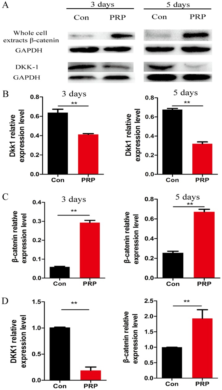
Platelet-rich plasma (PRP) is used in the clinic as an autologous blood product to stimulate bone regeneration and chondrogenesis. Numerous studies have demonstrated that PRP affects bone remodeling by accelerating osteoblast formation. With the research perspective focusing on osteoclasts, the present study established a mouse model of mandibular advancement to examine the effect of PRP on osteoclast differentiation induced by modification of the dynamics of the temporomandibular joint (TMJ). The lower incisors of the mice were trimmed by 1 mm and the resultant change in mandibular position during the process of eating induced condylar adaptation to this change. PRP significantly increased the bone mass and decreased osteoclastic activity, in vitro as well as in vivo. Mechanistically, the reduced expression of receptor activator of nuclear factor-κB ligand (RANKL)‑induced differentiation marker genes, including nuclear factor of activated T-cells, cytoplasmic 1, c-fos and tartrate-resistant acid phosphatase, and that of the resorptive activity marker genes such as cathepsin k, carbonic anhydrase 2 and matrix metalloproteinase 9, indicated that PRP suppresses RANKL-induced osteoclast differentiation. A microarray analysis revealed that several genes associated with the Wnt pathway were differentially expressed, which indicated the involvement of this pathway in osteoclast differentiation. Furthermore, the activation of the Wnt pathway was verified by reverse transcription-quantitative polymerase chain reaction and immunoblot analysis of Dickkopf-related protein 1 and β-catenin. The results of the present study indicated that PRP inhibits osteoclast differentiation through activation of the Wnt pathway.
富血小板血浆 (PRP) 作为一种自体血液产品,被临床用于刺激骨再生和软骨生成。大量研究表明,PRP 通过加速成骨细胞的形成来影响骨重塑。目前的研究以破骨细胞为研究视角,建立了下颌前伸的小鼠模型,以研究 PRP 对颞下颌关节 (TMJ) 动力学改变诱导的破骨细胞分化的影响。通过 1mm 修剪小鼠的下切牙,在进食过程中下颌位置的变化导致髁突适应这种变化。PRP 显著增加了骨量,降低了体外和体内的破骨细胞活性。从机制上讲,核因子-κB 受体激活剂配体 (RANKL) 诱导分化标记基因,包括激活 T 细胞的核因子、细胞质 1、c-fos 和抗酒石酸酸性磷酸酶的表达减少,以及吸收活性标记基因,如组织蛋白酶 K、碳酸酐酶 2 和基质金属蛋白酶 9 的表达减少,表明 PRP 抑制了 RANKL 诱导的破骨细胞分化。微阵列分析显示,几个与 Wnt 途径相关的基因表达存在差异,这表明该途径参与了破骨细胞分化。此外,通过 Dickkopf 相关蛋白 1 和 β-连环蛋白的逆转录定量聚合酶链反应和免疫印迹分析验证了 Wnt 途径的激活。本研究结果表明,PRP 通过激活 Wnt 途径抑制破骨细胞分化。