Miao Chenkui, Liang Chao, Tian Ye, Xu Aiming, Zhu Jundong, Zhao Kai, Zhang Jianzhong, Hua Yibo, Liu Shouyong, Dong Huiyu, Zhang Chao, Su Shifeng, Li Pu, Qin Chao, Wang Zengjun
State Key Laboratory of Reproductive Medicine and Department of Urology, The First Affiliated Hospital of Nanjing Medical University, Nanjing, China.
Department of Urology, Nanjing First Hospital, Nanjing Medical University, Nanjing, China.
Oncotarget. 2017 Oct 26;8(58):97811-97821. doi: 10.18632/oncotarget.22083. eCollection 2017 Nov 17.
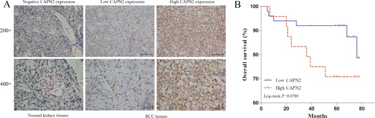
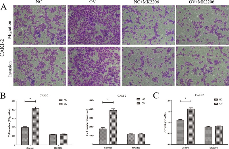
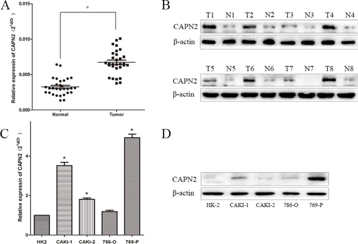
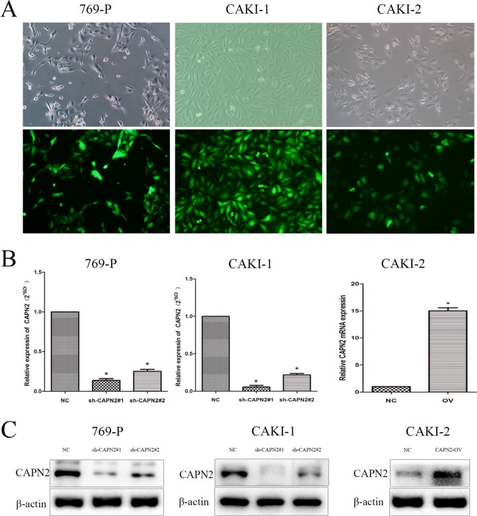
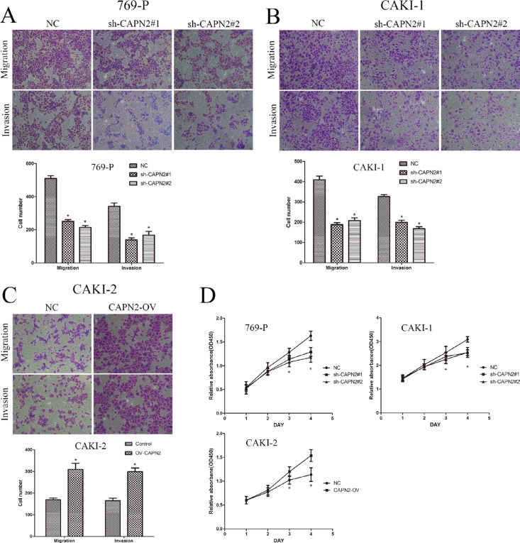
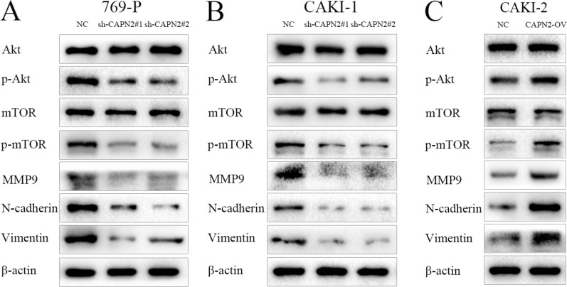
The calpain 2 (CAPN2) is upregulated in various malignant carcinomas. Previous studies have reported that CAPN2 functioned as an oncogenic factor in human cancers. However, its clinical role and potential effects on cell metastasis and proliferation in renal cell carcinoma (RCC) remain unknown. In this study, we evaluated the mRNA and protein levels of CAPN2 in human RCC specimens, matched normal specimens, and RCC cell lines using quantitative Real-time PCR (RT-PCR) and western blot. Immunohistochemistry of 74 RCC tissues in a tissue microarrays (TMAs) and normal kidney tissues were performed. Kaplan-Meier survival curve analyses were conducted to measure the correlation between CAPN2 and tumor prognosis. Cell migration, invasion and proliferation were detected by transwell assays and Cell Counting Kit-8 (CCK-8) assays. CAPN2 exhibited a significant overexpression in human RCC tissues and cell lines compared with adjacent non-tumor tissues and normal human proximal tubule epithelial cell line HK-2. Strong staining of CAPN2 was associated with higher clinical stage and histological grade. In addition, sh-CAPN2 could significantly inhibit migration, invasion and proliferation of 769-P and CAKI-1 cells. Conversely, increased cell biological behaviors were observed in CAPN2-OV CAKI-2 cells. Moreover, the subsequent mechanism investigation suggested that CAPN2 promoted tumor progression by activating AKT/mTOR signaling, enhancing epithelial mesenchymal transition (EMT) and MMP9 levels. The present study indicates that CAPN2 may act as a prominent indicator for RCC progression and a novel therapeutic target for RCC patients.
钙蛋白酶2(CAPN2)在多种恶性肿瘤中表达上调。先前的研究报道,CAPN2在人类癌症中作为一种致癌因子发挥作用。然而,其在肾细胞癌(RCC)中的临床作用以及对细胞转移和增殖的潜在影响仍不清楚。在本研究中,我们使用定量实时聚合酶链反应(RT-PCR)和蛋白质免疫印迹法评估了CAPN2在人RCC标本、配对的正常标本以及RCC细胞系中的mRNA和蛋白质水平。对组织芯片(TMAs)中的74例RCC组织和正常肾组织进行了免疫组织化学检测。进行了Kaplan-Meier生存曲线分析以衡量CAPN2与肿瘤预后之间的相关性。通过Transwell实验和细胞计数试剂盒-8(CCK-8)实验检测细胞迁移、侵袭和增殖情况。与相邻的非肿瘤组织和正常人近端肾小管上皮细胞系HK-2相比,CAPN2在人RCC组织和细胞系中显著过表达。CAPN2的强染色与更高的临床分期和组织学分级相关。此外,sh-CAPN2可显著抑制769-P和CAKI-1细胞的迁移、侵袭和增殖。相反,在CAPN2过表达的CAKI-2细胞中观察到细胞生物学行为增加。此外,后续的机制研究表明,CAPN2通过激活AKT/mTOR信号通路、增强上皮-间质转化(EMT)和MMP9水平来促进肿瘤进展。本研究表明,CAPN2可能是RCC进展的一个重要指标,也是RCC患者的一个新的治疗靶点。