Li Bobby W S, Stadhouders Ralph, de Bruijn Marjolein J W, Lukkes Melanie, Beerens Dior M J M, Brem Maarten D, KleinJan Alex, Bergen Ingrid, Vroman Heleen, Kool Mirjam, van IJcken Wilfred F J, Rao Tata Nageswara, Fehling Hans Jörg, Hendriks Rudi W
Department of Pulmonary Medicine, Rotterdam, Netherlands.
Center for Biomics, Erasmus MC Rotterdam, Rotterdam, Netherlands.
Front Immunol. 2017 Dec 1;8:1684. doi: 10.3389/fimmu.2017.01684. eCollection 2017.
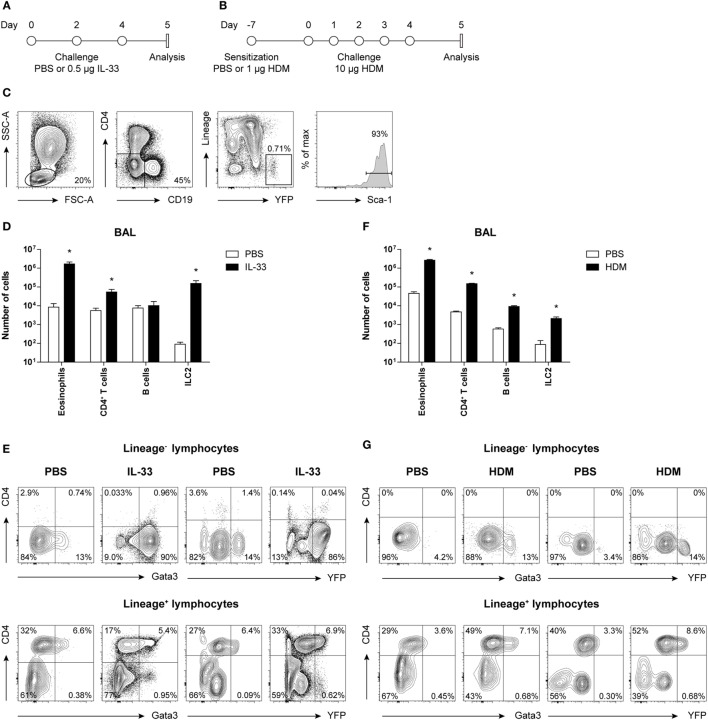
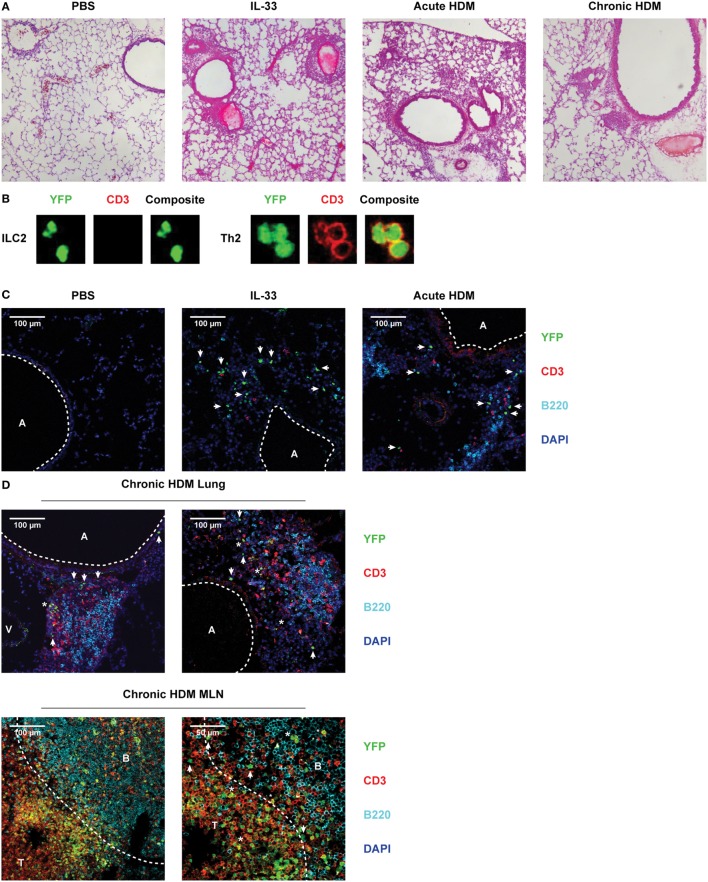
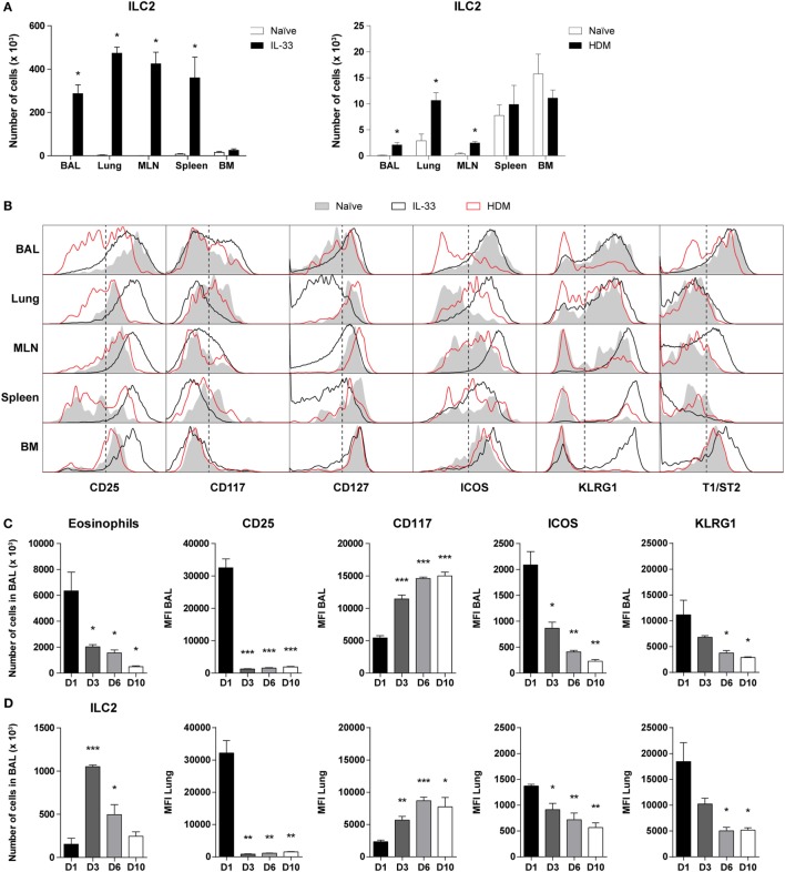
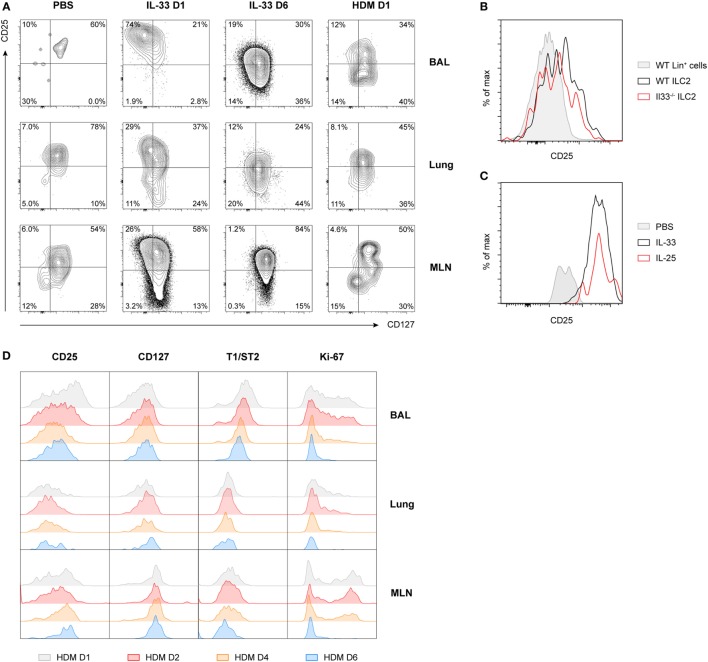
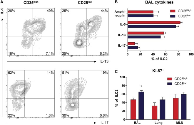
Group 2 innate lymphoid cells (ILC2) are implicated in allergic asthma as an early innate source of the type 2 cytokines IL-5 and IL-13. However, their induction in house dust mite (HDM)-mediated airway inflammation additionally requires T cell activation. It is currently unknown whether phenotypic differences exist between ILC2s that are activated in a T cell-dependent or T cell-independent fashion. Here, we compared ILC2s in IL-33- and HDM-driven airway inflammation. Using flow cytometry, we found that surface expression levels of various markers frequently used to identify ILC2s were dependent on their mode of activation, highly variable over time, and differed between tissue compartments, including bronchoalveolar lavage (BAL) fluid, lung, draining lymph nodes, and spleen. Whereas IL-33-activated BAL fluid ILC2s exhibited an almost uniform CD25CD127T1/ST2ICOSKLRG1 phenotype, at a comparable time point after HDM exposure BAL fluid ILC2s had a very heterogeneous surface marker phenotype. A major fraction of HDM-activated ILC2s were CD25CD127T1/ST2 ICOSKLRG1, but nevertheless had the capacity to produce large amounts of type 2 cytokines. HDM-activated CD25 ILC2s in BAL fluid and lung rapidly reverted to CD25 ILC2s upon stimulation with IL-33. Genome-wide transcriptional profiling of BAL ILC2s revealed ~1,600 differentially expressed genes: HDM-stimulated ILC2s specifically expressed genes involved in the regulation of adaptive immunity through B and T cell interactions, whereas IL-33-stimulated ILC2s expressed high levels of proliferation-related and cytokine genes. In both airway inflammation models ILC2s were present in the lung submucosa close to epithelial cells, as identified by confocal microscopy. In chronic HDM-driven airway inflammation ILC2s were also found inside organized cellular infiltrates near T cells. Collectively, our findings show that ILC2s are phenotypically more heterogeneous than previously thought, whereby their surface marker and gene expression profile are highly dynamic.
第2组固有淋巴细胞(ILC2)作为2型细胞因子IL-5和IL-13的早期固有来源,与过敏性哮喘有关。然而,它们在屋尘螨(HDM)介导的气道炎症中的诱导还需要T细胞活化。目前尚不清楚以T细胞依赖性或T细胞非依赖性方式活化的ILC2之间是否存在表型差异。在此,我们比较了IL-33和HDM驱动的气道炎症中的ILC2。使用流式细胞术,我们发现常用于鉴定ILC2的各种标志物的表面表达水平取决于它们的活化模式,随时间高度可变,并且在包括支气管肺泡灌洗(BAL)液、肺、引流淋巴结和脾脏在内的组织区室之间存在差异。虽然IL-33活化的BAL液ILC2表现出几乎一致的CD25⁺CD127⁺T1/ST2⁺ICOS⁻KLRG1⁻表型,但在HDM暴露后的可比时间点,BAL液ILC2具有非常异质的表面标志物表型。HDM活化的ILC2的很大一部分是CD25⁻CD127⁺T1/ST2⁺ICOS⁻KLRG1⁻,但仍有能力产生大量的2型细胞因子。BAL液和肺中HDM活化的CD25⁻ ILC2在用IL-33刺激后迅速恢复为CD25⁺ ILC2。BAL ILC2的全基因组转录谱分析揭示了约1600个差异表达基因:HDM刺激的ILC2特异性表达通过B和T细胞相互作用参与适应性免疫调节的基因,而IL-刺激的ILC2表达高水平的增殖相关基因和细胞因子基因。通过共聚焦显微镜鉴定,在两种气道炎症模型中,ILC2都存在于靠近上皮细胞的肺黏膜下层。在慢性HDM驱动的气道炎症中,在T细胞附近有组织的细胞浸润内也发现了ILC2。总的来说,我们的研究结果表明,ILC2在表型上比以前认为的更加异质,其表面标志物和基因表达谱具有高度动态性。