1 R&D Institute, Biosolution Inc., Seoul, South Korea.
2 Graduate School of Biotechnology, College of Life Science, Kyung Hee University, Yongin, South Korea.
Cell Transplant. 2017 Oct;26(10):1673-1687. doi: 10.1177/0963689717724794.
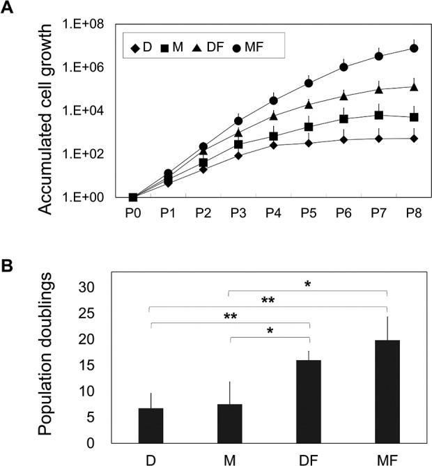
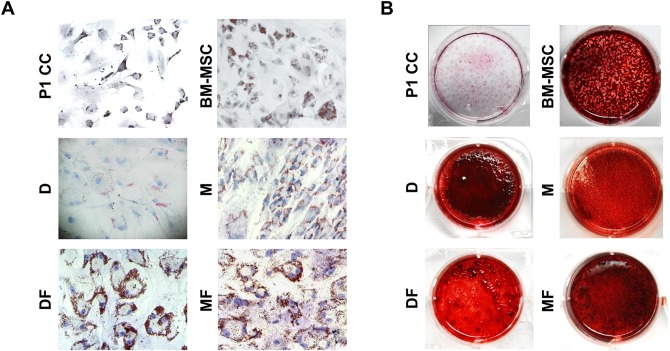
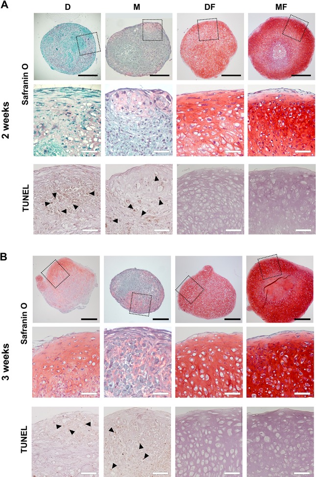
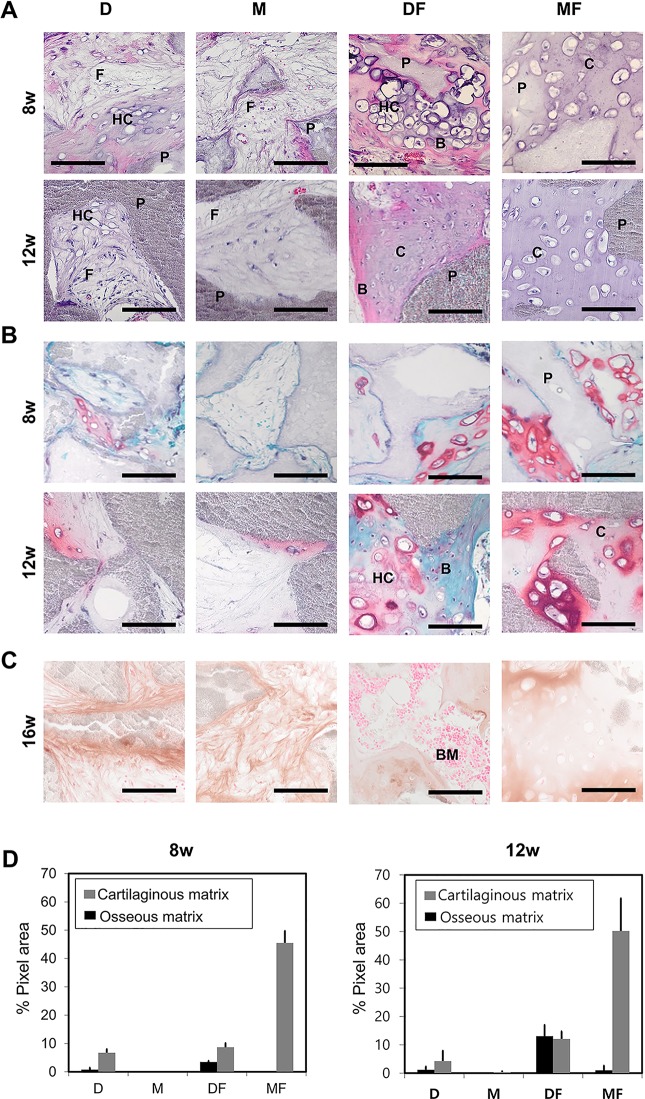
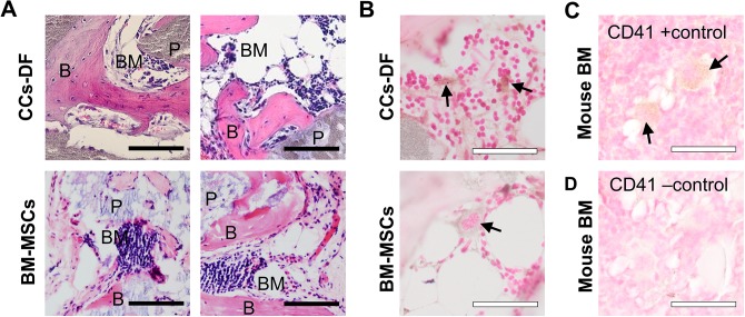
Given recent progress in regenerative medicine, we need a means to expand chondrocytes in quantity without losing their regenerative capability. Although many reports have shown that growth factor supplementation can have beneficial effects, the use of growth factor-supplemented basal media has widespread effect on the characteristics of chondrocytes. Chondrocytes were in vitro cultured in the 2 most widely used chondrocyte growth media, conventional chondrocyte culture medium and mesenchymal stem cell (MSC) culture medium, both with and without fibroblast growth factor-2 (FGF2) supplementation. Their expansion rates, expressions of extracellular matrix-related factors, senescence, and differentiation potentials were examined in vitro and in vivo. Our results revealed that chondrocytes quickly dedifferentiated during expansion in all tested media, as assessed by the loss of type II collagen expression. The 2 basal media (chondrocyte culture medium vs. MSC culture medium) were associated with distinct differences in cell senescence. Consistent with the literature, FGF2 was associated with accelerated dedifferentiation during expansion culture and superior redifferentiation upon induction. However, chondrocytes expanded in FGF2-containing conventional chondrocyte culture medium showed MSC-like features, as indicated by their ability to direct ectopic bone formation and cartilage formation. In contrast, chondrocytes cultured in FGF2-supplemented MSC culture medium showed potent chondrogenesis and almost no bone formation. The present findings show that the chosen basal medium can exert profound effects on the characteristics and activity of in vitro-expanded chondrocytes and indicate that right growth factor/medium combination can help chondrocytes retain a high-level chondrogenic potential without undergoing hypertrophic transition.
鉴于再生医学的最新进展,我们需要一种方法在不丧失其再生能力的情况下大量扩增软骨细胞。虽然许多报告表明生长因子补充可以产生有益的效果,但生长因子补充的基础培养基的使用对软骨细胞的特性有广泛的影响。将软骨细胞在两种最广泛使用的软骨细胞生长培养基(传统软骨细胞培养基和间充质干细胞(MSC)培养基)中进行体外培养,无论是否添加成纤维细胞生长因子 2(FGF2)。在体外和体内检查它们的扩增率、细胞外基质相关因子的表达、衰老和分化潜能。我们的结果表明,在所有测试的培养基中,软骨细胞在扩增过程中迅速去分化,这可以通过 II 型胶原蛋白表达的丧失来评估。两种基础培养基(软骨细胞培养基与 MSC 培养基)与细胞衰老之间存在明显差异。与文献一致,FGF2 在扩增培养过程中与加速去分化有关,并在诱导时具有更好的再分化能力。然而,在含有 FGF2 的传统软骨细胞培养基中扩增的软骨细胞表现出 MSC 样特征,这表明它们具有引导异位骨形成和软骨形成的能力。相比之下,在补充 FGF2 的 MSC 培养基中培养的软骨细胞表现出强大的软骨生成能力,几乎没有骨形成。这些发现表明,所选的基础培养基可以对体外扩增的软骨细胞的特性和活性产生深远影响,并表明正确的生长因子/培养基组合可以帮助软骨细胞保持高水平的软骨形成潜能,而不会发生肥大转化。