Xu Jin, Xu Yaozeng
Department of Orthopedics, The First Affiliated Hospital of Soochow University, No. 899 Ping Hai Road, Gusu District, Suzhou, 215031 China.
Department of Orthopedics, Baoshan District Shanghai Integrated Traditional Chinese and Western Medicine Hospital, Shanghai, 201999 China.
Cell Biosci. 2017 Dec 13;7:69. doi: 10.1186/s13578-017-0195-x. eCollection 2017.
Osteoarthritis (OA) is a chronic joint disease and there is no a definitive cure at present. Long non-coding RNAs (lncRNAs) have been confirmed to play important roles in the development of OA. However, the underlying mechanism of lncRNA maternally expressed gene 3 (MEG3) in OA has not been well elucidated.
The rat OA model and interleukin-1β (IL-1β)-induced rat chondrocytes were constructed. The expression pattern of lncRNA MEG3 and miR-16 was detected by RT-qPCR assay in cartilage tissues of rat OA model. The effect of MEG3 and miR-16 on IL-1β-induced chondrocytes was evaluated on the basis of cell viability and apoptosis. Then, the interaction among MEG3, miR-16 SMAD7 was explored by dual-luciferase reporter assay and RIP assay.
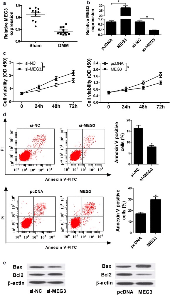
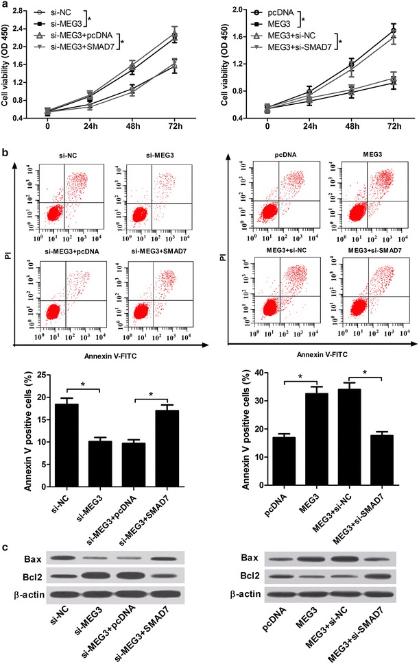
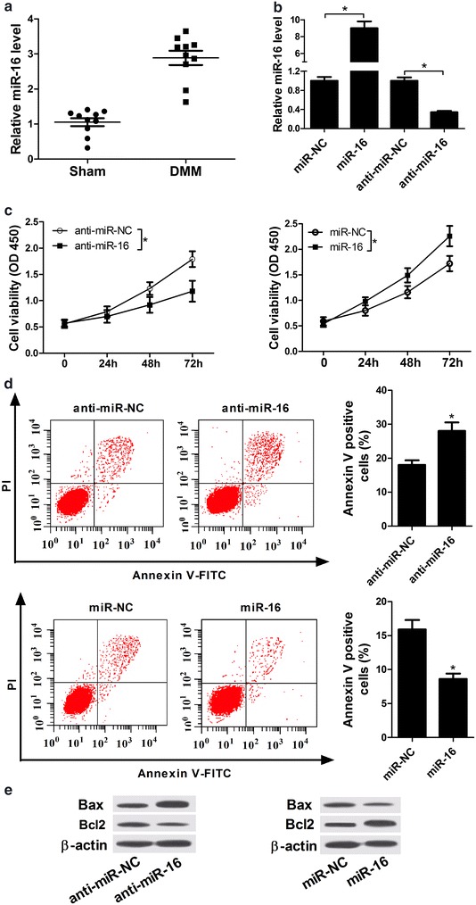
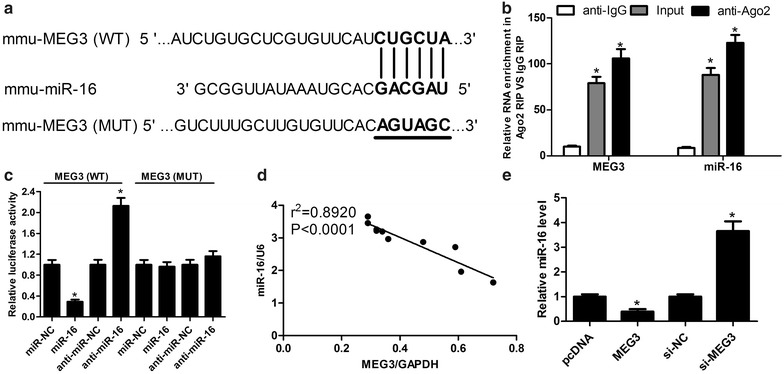
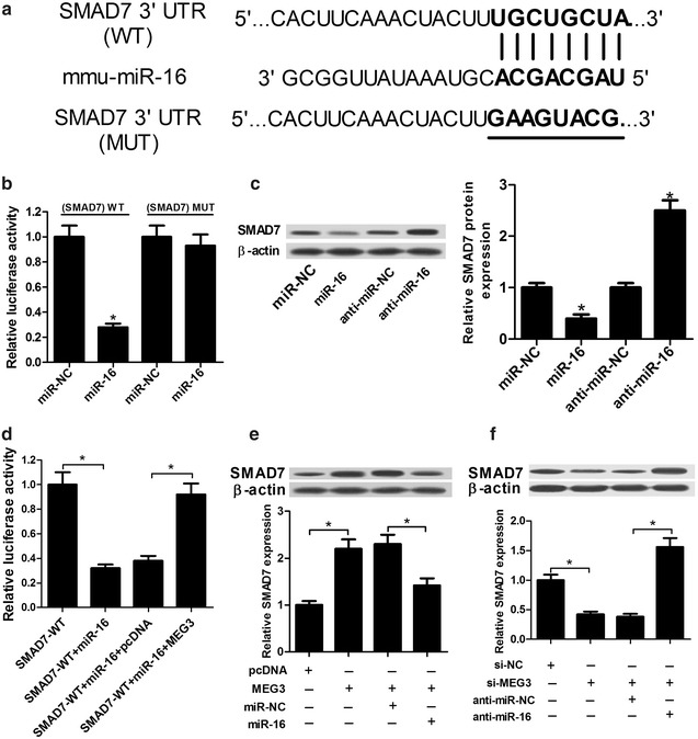
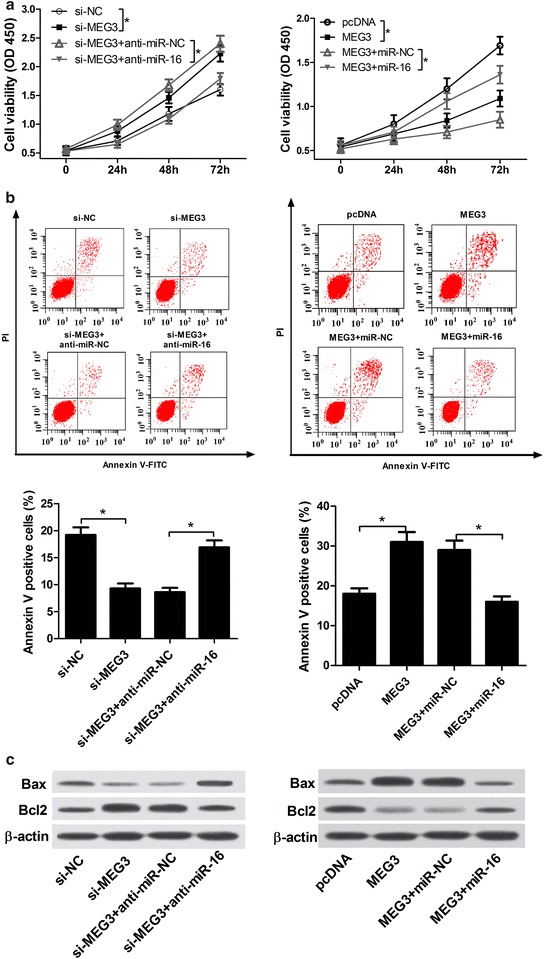
It is found that lncRNA MEG3 was down-regulated and miR-16 was up-regulated in rat OA cartilage tissues. MEG3 knockdown promoted proliferation and inhibited apoptosis, while miR-16 knockdown suppressed proliferation and promoted apoptosis in IL-1β-induced rat chondrocytes. Moreover, MEG3 was involved in miR-16 pathway and MEG3 suppressed miR-16 expression. Additionally, SMAD7 was a target gene of miR-16 and miR-16 suppressed SMAD7 expression in IL-1β-induced chondrocytes. Moreover, the expression of SMAD7 induced by MEG3 or si-MEG3 was markedly reversed by the introduction of miR-16 or anti-miR-16. Furthermore, MEG3 exerted its anti-proliferation and pro-apoptosis by regulating miR-16 and SMAD7.
MEG3 was down-regulated and miR-16 was up-regulated in cartilage tissues of rat OA model. MEG3 knockdown might lead to the progression of OA through miR-16/SMAD7 axis.
骨关节炎(OA)是一种慢性关节疾病,目前尚无确切的治愈方法。长链非编码RNA(lncRNAs)已被证实参与OA的发展过程。然而,lncRNA母系表达基因3(MEG3)在OA中的潜在机制尚未完全阐明。
构建大鼠OA模型和白细胞介素-1β(IL-1β)诱导的大鼠软骨细胞。采用RT-qPCR法检测大鼠OA模型软骨组织中lncRNA MEG3和miR-16的表达模式。基于细胞活力和凋亡情况,评估MEG3和miR-16对IL-1β诱导的软骨细胞的影响。然后,通过双荧光素酶报告基因检测和RNA免疫沉淀(RIP)检测探索MEG3、miR-16和SMAD7之间的相互作用。
发现lncRNA MEG3在大鼠OA软骨组织中表达下调,miR-1十六表达上调。敲低MEG3可促进IL-1β诱导的大鼠软骨细胞增殖并抑制其凋亡,而敲低miR-16则抑制增殖并促进凋亡。此外,MEG3参与miR-16通路,且MEG3抑制miR-16的表达。另外,SMAD7是miR-16的靶基因,miR-16在IL-1β诱导的软骨细胞中抑制SMAD7的表达。此外,引入miR-16或抗miR-16可显著逆转由MEG3或si-MEG3诱导的SMAD7的表达。此外,MEG3通过调节miR-16和SMAD7发挥其抗增殖和促凋亡作用。
在大鼠OA模型的软骨组织中,MEG3表达下调,miR-16表达上调。敲低MEG3可能通过miR-16/SMAD7轴导致OA进展。