Abu El-Asrar Ahmed M, Alam Kaiser, Garcia-Ramirez Marta, Ahmad Ajmal, Siddiquei Mohammad Mairaj, Mohammad Ghulam, Mousa Ahmed, De Hertogh Gert, Opdenakker Ghislain, Simó Rafael
Department of Ophthalmology, College of Medicine, King Saud University, Riyadh, Saudi Arabia.
Dr. Nasser Al-Rashid Research Chair in Ophthalmology, Riyadh, Saudi Arabia.
Mol Vis. 2017 Dec 5;23:853-871. eCollection 2017.
We investigated the link among the proinflammatory cytokine high-mobility group box 1 (HMGB1) and 8-hydroxy-2'-deoxyguanosine (8-OHdG) as a marker of oxidative DNA damage, the endothelial adhesion molecule and oxidase enzyme vascular adhesion protein-1 (VAP-1), and the inducible cytoprotective molecule heme oxygenase-1 (HO-1) in proliferative diabetic retinopathy (PDR). We correlated the levels of these molecules with clinical disease activity and studied the proinflammatory activities of HMGB1 on rat retinas and human retinal microvascular endothelial cells (HRMECs).
Vitreous samples from 47 PDR and 19 non-diabetic patients, epiretinal membranes from 11 patients with PDR, human retinas (16 from diabetic patients and 16 from non-diabetic subjects), rat retinas, and HRMECs were studied by enzyme-linked immunosorbent assay, immunohistochemistry, western blot immunofluorescence, and RT-PCR analyses. In addition, we assessed the adherence of leukocytes to HMGB1-stimulated HRMECs.
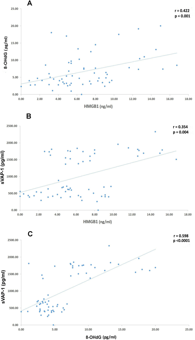
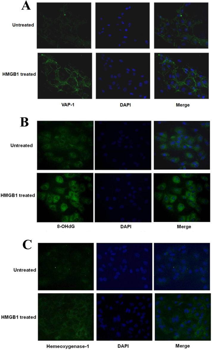
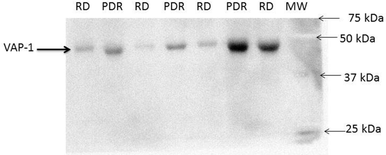
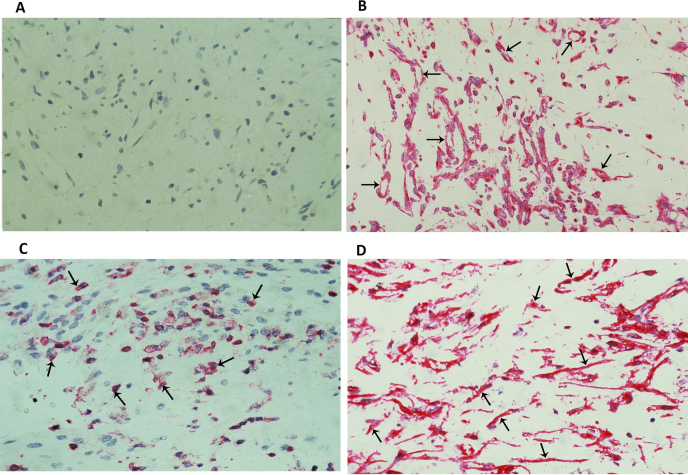
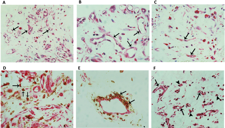
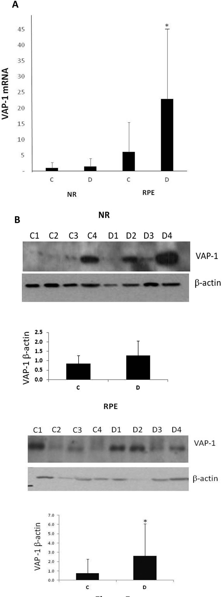
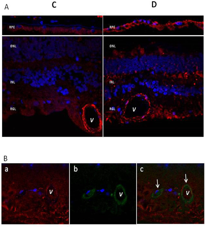
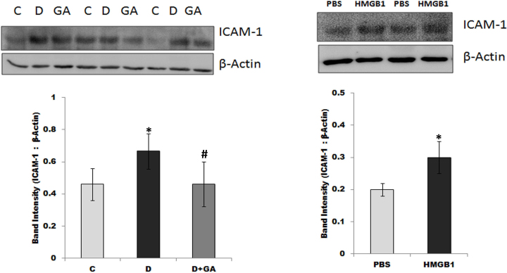
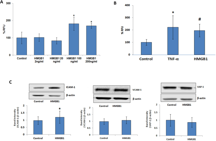
HMGB1, 8-OHdG, and soluble VAP-1 (sVAP-1) levels were significantly higher in vitreous samples from PDR patients than in those from non-diabetics (p = 0.001, <0.0001, <0.0001, respectively). The HMGB1, 8-OHdG, sVAP-1, and HO-1 levels in PDR with active neovascularization were significantly higher than those in inactive PDR (p = 0.025, <0.0001, <0.0001, 0.012, respectively). Significant positive correlations were observed between the levels of HMGB1 and the levels of 8-OHdG (r = 0.422; p = 0.001) and sVAP-1 (r = 0.354; p = 0.004) and between the levels of 8-OHdG and the levels of sVAP-1 (r = 0.598; p<0.0001). In epiretinal membranes, VAP-1 and 8-OHdG were expressed in vascular endothelial cells and stromal cells. Significant increases in the VAP-1 mRNA and protein levels were detected in the RPE, but not in the neuroretina of diabetic patients. Treatment of HRMEC with HMGB1, diabetes induction, and an intravitreal injection of HMGB1 in normal rats induced a significant upregulation of the adhesion molecule intercellular adhesion molecule-1 (ICAM-1) in HRMECs and retinas. On the other hand, the expressions of vascular cell adhesion molecule-1 and VAP-1 were not affected. Oral administration of the HMGB1 inhibitor glycyrrhizin in rats attenuated the diabetes-induced upregulation of the retinal ICAM-1 expression. Treatment of HRMECs with HMGB1 increased leukocyte adhesion and induced the upregulation of 8-OHdG and HO-1 and the membranous translocation of VAP-1.
Our results suggest a potential link among the proinflammatory cytokine HMGB1, VAP-1, oxidative stress, and HO-1 in the pathogenesis of PDR.
我们研究了促炎细胞因子高迁移率族蛋白B1(HMGB1)与作为氧化DNA损伤标志物的8-羟基-2'-脱氧鸟苷(8-OHdG)、内皮黏附分子及氧化酶血管黏附蛋白-1(VAP-1)以及增殖性糖尿病视网膜病变(PDR)中诱导性细胞保护分子血红素加氧酶-1(HO-1)之间的联系。我们将这些分子的水平与临床疾病活动度进行关联,并研究了HMGB1对大鼠视网膜和人视网膜微血管内皮细胞(HRMECs)的促炎活性。
通过酶联免疫吸附测定、免疫组织化学、蛋白质印迹免疫荧光和逆转录聚合酶链反应分析,对47例PDR患者和19例非糖尿病患者的玻璃体液样本、11例PDR患者的视网膜前膜、人视网膜(16例来自糖尿病患者,16例来自非糖尿病受试者)、大鼠视网膜和HRMECs进行研究。此外,我们评估了白细胞对HMGB1刺激的HRMECs的黏附情况。
PDR患者玻璃体液样本中的HMGB1、8-OHdG和可溶性VAP-1(sVAP-1)水平显著高于非糖尿病患者(分别为p = 0.001、<0.0001、<0.0001)。有活跃新生血管形成的PDR患者的HMGB1、8-OHdG、sVAP-1和HO-1水平显著高于无活跃病变的PDR患者(分别为p = 0.025、<0.0001、<0.0001、0.012)。观察到HMGB1水平与8-OHdG水平(r = 0.422;p = 0.001)以及sVAP-1水平(r = 0.354;p = 0.004)之间存在显著正相关,8-OHdG水平与sVAP-1水平之间也存在显著正相关(r = 0.598;p<0.0001)。在视网膜前膜中,VAP-1和8-OHdG在血管内皮细胞和基质细胞中表达。在糖尿病患者的视网膜色素上皮(RPE)中检测到VAP-1 mRNA和蛋白水平显著升高,但在神经视网膜中未检测到。用HMGB1处理HRMEC、诱导糖尿病以及在正常大鼠玻璃体内注射HMGB1均可诱导HRMECs和视网膜中黏附分子细胞间黏附分子-1(ICAM-