Tamura Ryo, Uemoto Shinji, Tabata Yasuhiko
Department of Surgery, Graduate School of Medicine, Kyoto University, Kyoto, Japan.
Department of Biomaterials, Field of Tissue Engineering, Institute for Frontier Medical Sciences, Kyoto University, Kyoto, Japan.
Inflamm Regen. 2016 Nov 17;36:26. doi: 10.1186/s41232-016-0030-5. eCollection 2016.
This study aimed to evaluate the effect of mesenchymal stem cell (MSC)-derived exosomes on an immune-induced liver injury model. MSCs show a unique function to modulate immune reaction although the molecular mechanisms are still under investigation. Exosomes are a nanoparticle containing microRNA and many ligands and are recognized as important factors secreted from MSC to express their function. This research is undertaken to evaluate the effect of MSC-derived exosome on concanavalin-A (con-A)-induced liver injury.
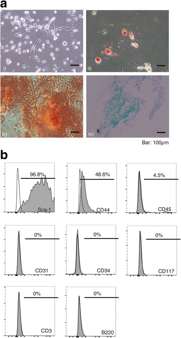
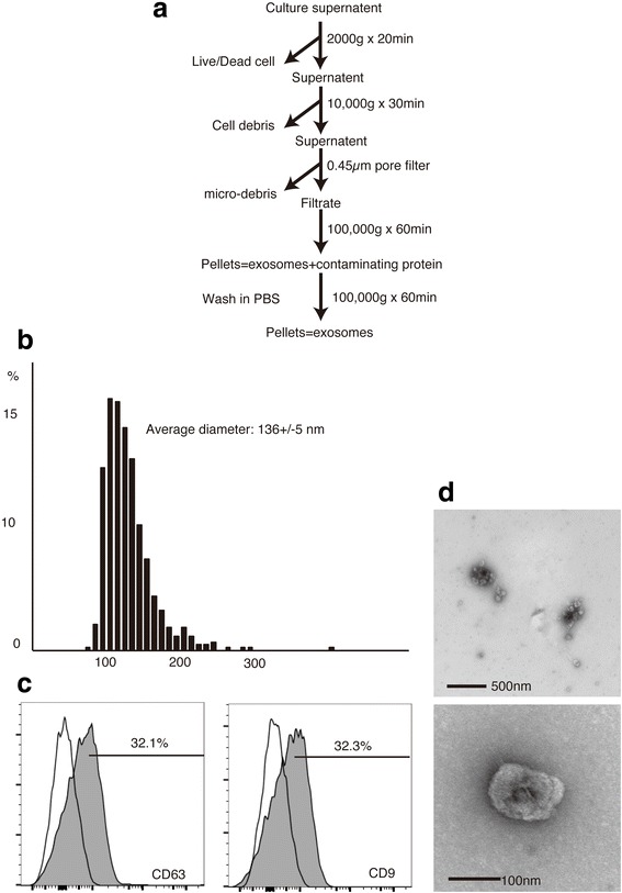
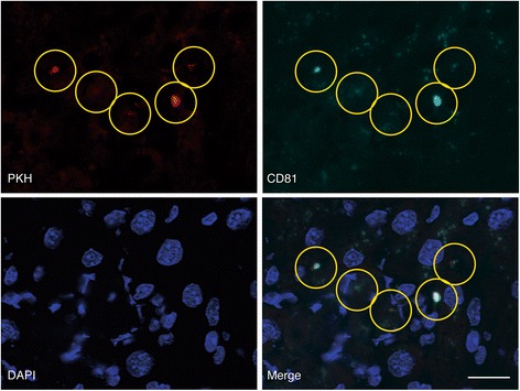
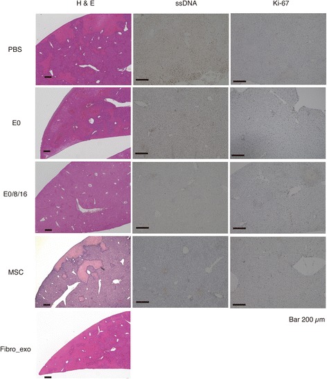
Exosomes were collected from the supernatant of MSC from the bone marrow of C57B6 mice with ultracentrifugation. The collected exosomes or MSCs were injected intravenously into liver injury mice that had been prepared by the intravenous con-A injection. Liver and serum samples were collected 24 h later to evaluate the macro- and microscopic images, the alanine aminotransferase (ALT), and cytokine messenger RNA (mRNA) expression levels. Phenotypical change of non-parenchymal liver cells was also evaluated by flow cytometry. Liver localization of PKH26 after the injection of PKH26-labeled exosomes or MSCs was observed by microscope. Each result was statistically analyzed with Student's test.
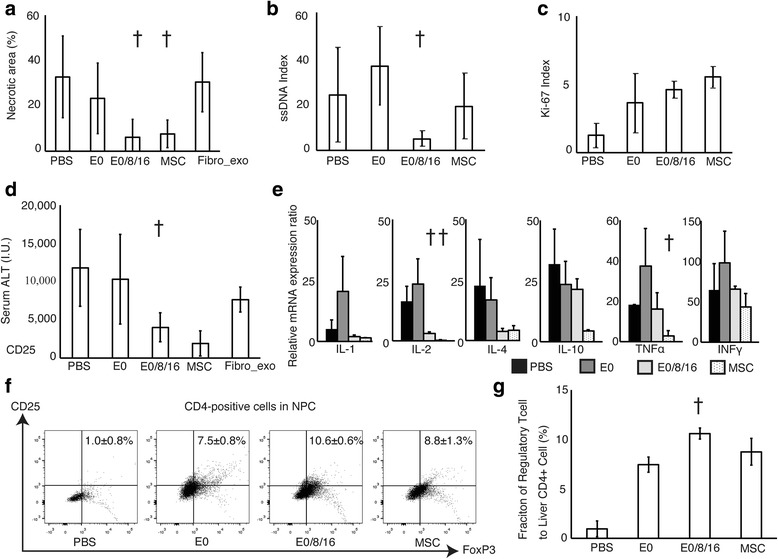
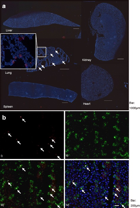
PKH was observed in the liver after PKH-labeled exosomes were injected into mouse, whereas it was only observed in the lung in a mouse group receiving PKH-leveled MSC. There were decreases in ALT, liver necrotic areas, and the extent of apoptosis indicated by the single-stranded DNA index of groups that received multiple injections of MSC-derived exosomes, but an increase in the Ki-67 index. The mRNA expression of anti-inflammatory cytokines was enhanced. The number of Treg was increased among NPCs in a group receiving exosomes multiple times.
Suppression of con-A-induced liver injury by injection of exosomes was observed as same extent as MSC. Considering the advantage of exosomes as its non-living nature and dosing adjustability over MSC, exosome will be one alternative of MSC transplantation.
本研究旨在评估间充质干细胞(MSC)来源的外泌体对免疫诱导性肝损伤模型的影响。尽管分子机制仍在研究中,但MSC显示出调节免疫反应的独特功能。外泌体是一种包含微小RNA和许多配体的纳米颗粒,被认为是MSC分泌以发挥其功能的重要因子。本研究旨在评估MSC来源的外泌体对刀豆蛋白A(Con-A)诱导的肝损伤的影响。
通过超速离心从C57B6小鼠骨髓的MSC上清液中收集外泌体。将收集的外泌体或MSC静脉注射到通过静脉注射Con-A制备的肝损伤小鼠中。24小时后收集肝脏和血清样本,以评估大体和微观图像、丙氨酸转氨酶(ALT)以及细胞因子信使核糖核酸(mRNA)表达水平。还通过流式细胞术评估非实质肝细胞的表型变化。通过显微镜观察注射PKH26标记的外泌体或MSC后PKH26在肝脏中的定位。每个结果均采用学生t检验进行统计学分析。
将PKH标记的外泌体注射到小鼠体内后,在肝脏中观察到PKH,而在接受PKH标记的MSC的小鼠组中仅在肺中观察到PKH。多次注射MSC来源外泌体的组中,ALT、肝坏死面积和由单链DNA指数表示的凋亡程度降低,但Ki-67指数升高。抗炎细胞因子的mRNA表达增强。多次接受外泌体的组中,非实质细胞中调节性T细胞(Treg)的数量增加。
观察到注射外泌体对Con-A诱导的肝损伤的抑制作用与MSC相同。考虑到外泌体相对于MSC的非活性性质和剂量可调节性的优势,外泌体将成为MSC移植的一种替代选择。