• 文献检索
  • 文档翻译
  • 深度研究
  • 学术资讯
  • Suppr Zotero 插件Zotero 插件
  • 邀请有礼
  • 套餐&价格
  • 历史记录
应用&插件
Suppr Zotero 插件Zotero 插件浏览器插件Mac 客户端Windows 客户端微信小程序
定价
高级版会员购买积分包购买API积分包
服务
文献检索文档翻译深度研究API 文档MCP 服务
关于我们
关于 Suppr公司介绍联系我们用户协议隐私条款
关注我们

Suppr 超能文献

核心技术专利:CN118964589B侵权必究
粤ICP备2023148730 号-1Suppr @ 2026

文献检索

告别复杂PubMed语法,用中文像聊天一样搜索,搜遍4000万医学文献。AI智能推荐,让科研检索更轻松。

立即免费搜索

文件翻译

保留排版,准确专业,支持PDF/Word/PPT等文件格式,支持 12+语言互译。

免费翻译文档

深度研究

AI帮你快速写综述,25分钟生成高质量综述,智能提取关键信息,辅助科研写作。

立即免费体验

多发性硬化症非视神经炎眼中进行性内核层功能障碍。

Progressive inner nuclear layer dysfunction in non-optic neuritis eyes in MS.

作者信息

You Yuyi, Graham Elizabeth C, Shen Ting, Yiannikas Con, Parratt John, Gupta Vivek, Barton Joshua, Dwyer Michael, Barnett Michael H, Fraser Clare L, Graham Stuart L, Klistorner Alexander

机构信息

Save Sight Institute (Y.Y., E.C.G., C.L.F., A.K.), The University of Sydney; Department of Health and Medical Sciences (Y.Y., T.S., V.G., S.L.G., A.K.), Macquarie University; Department of Neurology (C.Y., J.P.), Royal North Shore Hospital; Brain and Mind Center (J.B., M.H.B.), The University of Sydney; Sydney Neuroimaging Analysis Center (M.H.B., A.K.), New South Wales, Australia; and Buffalo Neuroimaging Analysis Center (M.D.), University at Buffalo, NY.

出版信息

Neurol Neuroimmunol Neuroinflamm. 2017 Dec 15;5(1):e427. doi: 10.1212/NXI.0000000000000427. eCollection 2018 Jan.

DOI:10.1212/NXI.0000000000000427
PMID:29259999
原文链接:https://pmc.ncbi.nlm.nih.gov/articles/PMC5732006/
Abstract

OBJECTIVE

To investigate primary retinal functional changes in non-optic neuritis (ON) eyes of patients with MS by full-field electroretinography (ERG).

METHODS

Seventy-seven patients with relapsing-remitting MS with no history of clinical ON in at least 1 eye and 30 healthy controls were recruited in the cohort study. Full-field ERGs were recorded, and retinal optical coherence tomography scans were performed to assess the thicknesses of peripapillary retinal nerve fiber layer (RNFL) and retinal ganglion cell layer-inner plexiform layer (GCL-IPL). Annual MRI scans were also carried out to evaluate the disease activity in the brain. Patients were followed up for 3 years.

RESULTS

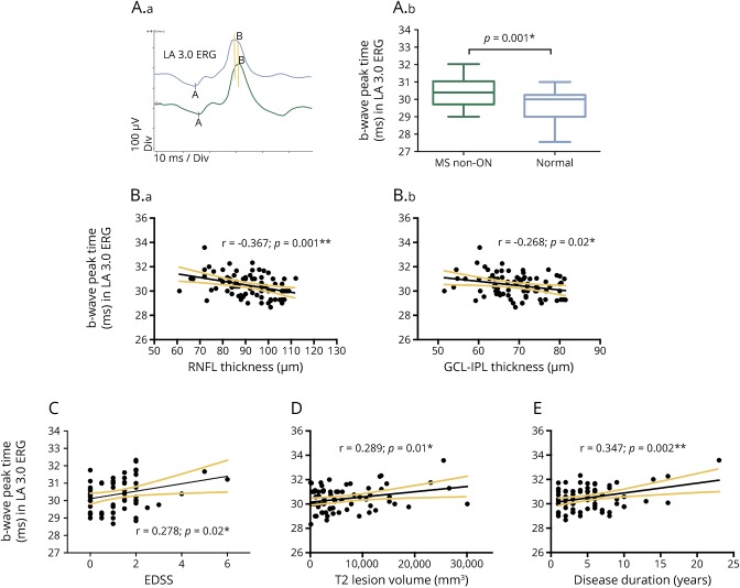
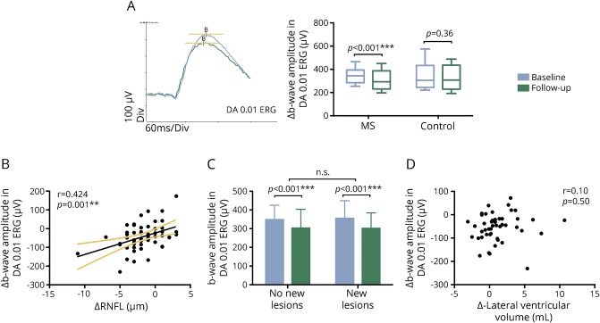
At baseline, a delayed b-wave peak time was observed in the cone response ( < 0.001), which was associated with the thicknesses of RNFL and GCL-IPL. The peak time of the delayed b-wave also correlated with the Expanded Disability Status Scale, T2 lesion volume, and disease duration. During the 3-year follow-up, progressive ERG amplitude reduction was observed (both a- and b-waves, < 0.05). There was a correlation between the b-wave amplitude reduction and longitudinal RNFL loss ( = 0.001). However, no correlation was found between longitudinal ERG changes and disease activity in the brain.

CONCLUSIONS

This study demonstrated progressive inner nuclear layer dysfunction in MS. The borderline a-wave changes suggested some outer retinal dysfunction as well. The correlation between full-field ERG changes and retinal ganglion cell loss suggested that there might be subclinical retinal pathology in MS affecting both outer and inner retinal layers.

摘要

目的

通过全视野视网膜电图(ERG)研究多发性硬化症(MS)患者非视神经炎(ON)眼的原发性视网膜功能变化。

方法

在队列研究中招募了77例复发缓解型MS患者,这些患者至少一只眼无临床ON病史,以及30名健康对照者。记录全视野ERG,并进行视网膜光学相干断层扫描以评估视乳头周围视网膜神经纤维层(RNFL)和视网膜神经节细胞层-内丛状层(GCL-IPL)的厚度。还进行年度MRI扫描以评估脑部疾病活动。对患者进行了3年的随访。

结果

在基线时,在视锥细胞反应中观察到b波峰时间延迟(<0.001),这与RNFL和GCL-IPL的厚度相关。延迟b波的峰时间也与扩展残疾状态量表、T2病变体积和病程相关。在3年随访期间,观察到ERG振幅逐渐降低(a波和b波,均<0.05)。b波振幅降低与纵向RNFL损失之间存在相关性(=0.001)。然而,未发现纵向ERG变化与脑部疾病活动之间存在相关性。

结论

本研究证明了MS患者存在进行性内核层功能障碍。临界a波变化也提示存在一些外层视网膜功能障碍。全视野ERG变化与视网膜神经节细胞丢失之间的相关性表明,MS可能存在亚临床视网膜病变,影响外层和内层视网膜。

https://cdn.ncbi.nlm.nih.gov/pmc/blobs/d42c/5732006/d710c6ecfcef/NEURIMMINFL2017014092f2.jpg
https://cdn.ncbi.nlm.nih.gov/pmc/blobs/d42c/5732006/aea7a61cb15f/NEURIMMINFL2017014092f1.jpg
https://cdn.ncbi.nlm.nih.gov/pmc/blobs/d42c/5732006/d710c6ecfcef/NEURIMMINFL2017014092f2.jpg
https://cdn.ncbi.nlm.nih.gov/pmc/blobs/d42c/5732006/aea7a61cb15f/NEURIMMINFL2017014092f1.jpg
https://cdn.ncbi.nlm.nih.gov/pmc/blobs/d42c/5732006/d710c6ecfcef/NEURIMMINFL2017014092f2.jpg

相似文献

1
Progressive inner nuclear layer dysfunction in non-optic neuritis eyes in MS.多发性硬化症非视神经炎眼中进行性内核层功能障碍。
Neurol Neuroimmunol Neuroinflamm. 2017 Dec 15;5(1):e427. doi: 10.1212/NXI.0000000000000427. eCollection 2018 Jan.
2
Ganglion cell loss in relation to visual disability in multiple sclerosis.多发性硬化症中与视力障碍相关的神经节细胞丢失。
Ophthalmology. 2012 Jun;119(6):1250-7. doi: 10.1016/j.ophtha.2011.11.032. Epub 2012 Feb 23.
3
Regional Analysis of Inner Retinal Layer Changes in Multiple Sclerosis with and without Optic Neuritis.多发性硬化症伴或不伴视神经炎的内视网膜层变化的区域分析。
Turk J Ophthalmol. 2023 Jun 21;53(3):169-174. doi: 10.4274/tjo.galenos.2023.81486.
4
Visual dysfunction in multiple sclerosis correlates better with optical coherence tomography derived estimates of macular ganglion cell layer thickness than peripapillary retinal nerve fiber layer thickness.多发性硬化症的视觉功能障碍与光学相干断层扫描测量的黄斑神经节细胞层厚度相关性优于视盘周围视网膜神经纤维层厚度。
Mult Scler. 2011 Dec;17(12):1449-63. doi: 10.1177/1352458511418630. Epub 2011 Aug 24.
5
Inner nuclear layer thickening is inversley proportional to retinal ganglion cell loss in optic neuritis.内核层增厚与视神经炎中视网膜神经节细胞丢失成反比。
PLoS One. 2013 Oct 3;8(10):e78341. doi: 10.1371/journal.pone.0078341. eCollection 2013.
6
Progressive Loss of Retinal Ganglion Cells and Axons in Nonoptic Neuritis Eyes in Multiple Sclerosis: A Longitudinal Optical Coherence Tomography Study.多发性硬化症非视神经炎眼中视网膜神经节细胞和轴突的渐进性丧失:一项纵向光学相干断层扫描研究
Invest Ophthalmol Vis Sci. 2016 Apr 1;57(4):2311-7. doi: 10.1167/iovs.15-19047.
7
Optical coherence tomography segmentation analysis in relapsing remitting versus progressive multiple sclerosis.复发缓解型与进展型多发性硬化症的光学相干断层扫描分割分析
PLoS One. 2017 Feb 13;12(2):e0172120. doi: 10.1371/journal.pone.0172120. eCollection 2017.
8
Anatomical effects of intravitreal anti-vascular endothelial growth factor injections on inner layers of the lesion-free retina.玻璃体腔抗血管内皮生长因子注射对无病变视网膜内层的解剖学影响。
Cutan Ocul Toxicol. 2021 Jun;40(2):135-139. doi: 10.1080/15569527.2021.1919136. Epub 2021 May 4.
9
Evaluation of the Innermost Retinal Layers and Visual Evoked Potentials in Patients with Multiple Sclerosis.多发性硬化症患者最内层视网膜层及视觉诱发电位的评估
Curr Eye Res. 2016 Oct;41(10):1353-1358. doi: 10.3109/02713683.2015.1119283. Epub 2016 Feb 16.
10
Determination of retinal nerve fibre layer and ganglion cell/inner plexiform layers progression rates using two optical coherence tomography systems: The PROGRESSA study.使用两种光学相干断层扫描系统测定视网膜神经纤维层和节细胞/内丛状层进展率:进展研究。
Clin Exp Ophthalmol. 2020 Sep;48(7):915-926. doi: 10.1111/ceo.13826. Epub 2020 Aug 14.

引用本文的文献

1
Retinal Function in Advanced Multiple Sclerosis.晚期多发性硬化症的视网膜功能。
Invest Ophthalmol Vis Sci. 2024 Nov 4;65(13):2. doi: 10.1167/iovs.65.13.2.
2
Assessing the visual afferent pathway with the multifocal visual evoked potentials in the radiologically isolated syndrome.评估影像学孤立综合征的多焦视觉诱发电位的视觉传入通路。
Sci Rep. 2024 Aug 30;14(1):20169. doi: 10.1038/s41598-024-68825-6.
3
Utility of the visual system to monitor neurodegeneration in multiple sclerosis.视觉系统在监测多发性硬化症神经退行性变中的作用。

本文引用的文献

1
Retinal layer segmentation in multiple sclerosis: a systematic review and meta-analysis.多发性硬化症中的视网膜层分割:系统评价和荟萃分析。
Lancet Neurol. 2017 Oct;16(10):797-812. doi: 10.1016/S1474-4422(17)30278-8. Epub 2017 Sep 12.
2
Neurological software tool for reliable atrophy measurement (NeuroSTREAM) of the lateral ventricles on clinical-quality T2-FLAIR MRI scans in multiple sclerosis.用于在多发性硬化症的临床质量 T2-FLAIR MRI 扫描上进行可靠的侧脑室萎缩测量的神经学软件工具 (NeuroSTREAM)。
Neuroimage Clin. 2017 Jun 19;15:769-779. doi: 10.1016/j.nicl.2017.06.022. eCollection 2017.
3
Optical coherence tomography segmentation analysis in relapsing remitting versus progressive multiple sclerosis.
Front Mol Neurosci. 2023 Mar 29;16:1125115. doi: 10.3389/fnmol.2023.1125115. eCollection 2023.
4
Treatment with MDL 72527 Ameliorated Clinical Symptoms, Retinal Ganglion Cell Loss, Optic Nerve Inflammation, and Improved Visual Acuity in an Experimental Model of Multiple Sclerosis.MDL 72527 治疗改善多发性硬化症实验模型中的临床症状、视网膜神经节细胞丢失、视神经炎症和提高视力。
Cells. 2022 Dec 16;11(24):4100. doi: 10.3390/cells11244100.
5
Structural and functional markers of optic nerve damage in myelin oligodendrocyte glycoprotein antibody-associated optic neuritis.髓鞘少突胶质细胞糖蛋白抗体相关视神经炎中视神经损伤的结构和功能标志物
Mult Scler J Exp Transl Clin. 2021 Dec 21;7(4):20552173211063126. doi: 10.1177/20552173211063126. eCollection 2021 Oct.
6
Optical coherence tomography in multiple sclerosis: A 3-year prospective multicenter study.光学相干断层扫描在多发性硬化症中的应用:一项为期 3 年的前瞻性多中心研究。
Ann Clin Transl Neurol. 2021 Dec;8(12):2235-2251. doi: 10.1002/acn3.51473. Epub 2021 Nov 18.
7
A three-year longitudinal study of retinal function and structure in patients with multiple sclerosis.一项针对多发性硬化症患者视网膜功能和结构的三年纵向研究。
Doc Ophthalmol. 2022 Feb;144(1):3-16. doi: 10.1007/s10633-021-09855-7. Epub 2021 Oct 27.
8
Normative Data of Axial Length, Retinal Thickness Measurements, Visual Evoked Potentials, and Full-Field Electroretinography in Female, Wild-Type Minipigs.女性野生型小型猪眼轴长度、视网膜厚度测量、视觉诱发电位和全视野视网膜电图的正常值。
Transl Vis Sci Technol. 2021 Oct 4;10(12):3. doi: 10.1167/tvst.10.12.3.
9
Novel Foveal Features Associated With Vision Impairment in Multiple Sclerosis.多发性硬化症相关视力损害的新黄斑特征。
Invest Ophthalmol Vis Sci. 2021 Sep 2;62(12):27. doi: 10.1167/iovs.62.12.27.
10
Disturbed Presynaptic Ca Signaling in Photoreceptors in the EAE Mouse Model of Multiple Sclerosis.多发性硬化症实验性自身免疫性脑脊髓炎(EAE)小鼠模型中光感受器突触前钙信号传导紊乱
iScience. 2020 Nov 20;23(12):101830. doi: 10.1016/j.isci.2020.101830. eCollection 2020 Dec 18.
复发缓解型与进展型多发性硬化症的光学相干断层扫描分割分析
PLoS One. 2017 Feb 13;12(2):e0172120. doi: 10.1371/journal.pone.0172120. eCollection 2017.
4
Age Related Multiple Sclerosis Severity Score: Disability ranked by age.年龄相关多发性硬化严重程度评分:按年龄分级的残疾程度。
Mult Scler. 2017 Dec;23(14):1938-1946. doi: 10.1177/1352458517690618. Epub 2017 Feb 3.
5
Progressive Loss of Retinal Ganglion Cells and Axons in Nonoptic Neuritis Eyes in Multiple Sclerosis: A Longitudinal Optical Coherence Tomography Study.多发性硬化症非视神经炎眼中视网膜神经节细胞和轴突的渐进性丧失:一项纵向光学相干断层扫描研究
Invest Ophthalmol Vis Sci. 2016 Apr 1;57(4):2311-7. doi: 10.1167/iovs.15-19047.
6
Retinal thickness measured with optical coherence tomography and risk of disability worsening in multiple sclerosis: a cohort study.光学相干断层扫描测量的视网膜厚度与多发性硬化症残疾恶化风险:一项队列研究。
Lancet Neurol. 2016 May;15(6):574-84. doi: 10.1016/S1474-4422(16)00068-5. Epub 2016 Mar 18.
7
Correlation between brain volume loss and clinical and MRI outcomes in multiple sclerosis.多发性硬化症中脑容量损失与临床及磁共振成像结果之间的相关性。
Neurology. 2015 Feb 24;84(8):784-93. doi: 10.1212/WNL.0000000000001281. Epub 2015 Jan 28.
8
Axonal loss of retinal neurons in multiple sclerosis associated with optic radiation lesions.多发性硬化症伴视辐射病变时视网膜神经元的轴突丧失。
Neurology. 2014 Jun 17;82(24):2165-72. doi: 10.1212/WNL.0000000000000522. Epub 2014 May 16.
9
Tracking changes over time in retinal nerve fiber layer and ganglion cell-inner plexiform layer thickness in multiple sclerosis.追踪多发性硬化症患者视网膜神经纤维层和神经节细胞-内丛状层厚度随时间的变化
Mult Scler. 2014 Sep;20(10):1331-41. doi: 10.1177/1352458514523498. Epub 2014 Mar 17.
10
Mechanisms of neurodegeneration and axonal dysfunction in multiple sclerosis.多发性硬化症中的神经退行性变和轴突功能障碍的机制。
Nat Rev Neurol. 2014 Apr;10(4):225-38. doi: 10.1038/nrneurol.2014.37. Epub 2014 Mar 18.