NeuroCure Clinical Research Center and Experimental and Clinical Research Center, Max Delbrueck Center for Molecular Medicine and Charité - Universitätsmedizin Berlin, Berlin, Germany.
Department of Neurology, Johns Hopkins Hospital, Baltimore, Maryland, USA.
Ann Clin Transl Neurol. 2021 Dec;8(12):2235-2251. doi: 10.1002/acn3.51473. Epub 2021 Nov 18.
To evaluate changes over 3 years in the thickness of inner retinal layers including the peripapillary retinal nerve fiber layer (pRNFL), and combined macular ganglion cell and inner plexiform layers (mGCIPL), in individuals with relapsing-remitting multiple sclerosis (RRMS) versus healthy controls; to determine whether optical coherence tomography (OCT) is sufficiently sensitive and reproducible to detect small degrees of neuroaxonal loss over time that correlate with changes in brain volume and disability progression as measured by the Expanded Disability Status Scale (EDSS).
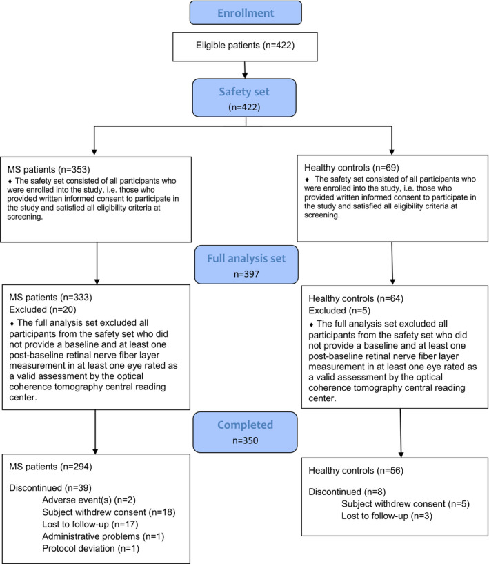
Individuals with RRMS from 28 centers (n = 333) were matched with 64 healthy participants. OCT scans were performed on Heidelberg Spectralis machines (at baseline; 1 month; 6 months; 6-monthly thereafter).
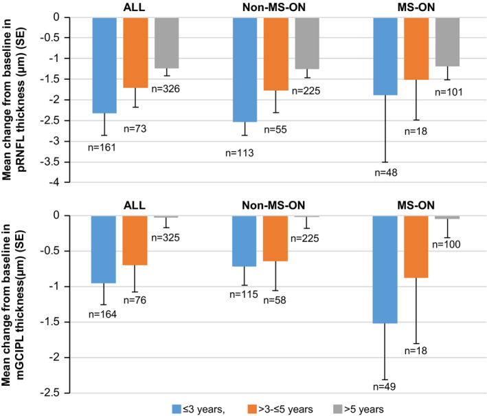
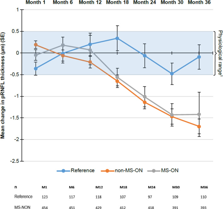
OCT measurements were highly reproducible between baseline and 1 month (intraclass correlation coefficient >0.98). Significant inner retinal layer thinning was observed in individuals with multiple sclerosis (MS) compared with controls regardless of previous MS-associated optic neuritis--group differences (95% CI) over 3 years: pRNFL: -1.86 (-2.54, -1.17) µm; mGCIPL: -2.03 (-2.78, -1.28) µm (both p < 0.0001; effect sizes 0.39 and 0.34). Greater inner retinal layer atrophy was observed in individuals diagnosed with RRMS <3 years versus >5 years (pRNFL: p < 0.05; mGCIPL: p < 0.01). Brain volume decreased by 1.3% in individuals with MS over 3 years compared to 0.5% in control subjects (effect size 0.76). mGCIPL atrophy correlated with brain atrophy (p < 0.0001). There was no correlation of OCT data with disability progression.
OCT has potential to estimate rates of neurodegeneration in the retina and brain. The effect size for OCT, smaller than for magnetic resonance imaging based on Heidelberg Spectralis data acquired in this study, was increased in early disease.
评估 3 年内包括视盘周围视网膜神经纤维层(pRNFL)和联合黄斑神经节细胞和内丛状层(mGCIPL)在内的内视网膜层厚度在复发缓解型多发性硬化症(RRMS)患者与健康对照者中的变化;确定光学相干断层扫描(OCT)是否足够敏感和可重复,以检测随时间推移与脑体积变化和扩展残疾状况量表(EDSS)测量的残疾进展相关的较小程度的神经轴突丢失。
来自 28 个中心的 RRMS 患者(n=333)与 64 名健康参与者相匹配。在海德堡 Spectralis 机器上进行 OCT 扫描(基线;1 个月;6 个月;此后每 6 个月一次)。
基线和 1 个月之间的 OCT 测量值高度可重复(组内相关系数>0.98)。与对照组相比,多发性硬化症患者的内视网膜层明显变薄,无论是否存在先前的多发性硬化症相关视神经炎——3 年内的组间差异(95%置信区间):pRNFL:-1.86(-2.54,-1.17)μm;mGCIPL:-2.03(-2.78,-1.28)μm(均p<0.0001;效应大小 0.39 和 0.34)。RRMS 诊断时间<3 年的患者比>5 年的患者观察到更大的内视网膜层萎缩(pRNFL:p<0.05;mGCIPL:p<0.01)。与对照组相比,多发性硬化症患者在 3 年内脑体积减少 1.3%,而对照组为 0.5%(效应大小 0.76)。mGCIPL 萎缩与脑萎缩相关(p<0.0001)。OCT 数据与残疾进展无相关性。
OCT 有可能估计视网膜和大脑中的神经退行性变速度。基于本研究中使用海德堡 Spectralis 数据获取的磁共振成像,OCT 的效应大小较小,但在早期疾病中增加。