• 文献检索
  • 文档翻译
  • 深度研究
  • 学术资讯
  • Suppr Zotero 插件Zotero 插件
  • 邀请有礼
  • 套餐&价格
  • 历史记录
应用&插件
Suppr Zotero 插件Zotero 插件浏览器插件Mac 客户端Windows 客户端微信小程序
定价
高级版会员购买积分包购买API积分包
服务
文献检索文档翻译深度研究API 文档MCP 服务
关于我们
关于 Suppr公司介绍联系我们用户协议隐私条款
关注我们

Suppr 超能文献

核心技术专利:CN118964589B侵权必究
粤ICP备2023148730 号-1Suppr @ 2026

文献检索

告别复杂PubMed语法,用中文像聊天一样搜索,搜遍4000万医学文献。AI智能推荐,让科研检索更轻松。

立即免费搜索

文件翻译

保留排版,准确专业,支持PDF/Word/PPT等文件格式,支持 12+语言互译。

免费翻译文档

深度研究

AI帮你快速写综述,25分钟生成高质量综述,智能提取关键信息,辅助科研写作。

立即免费体验

晚期多发性硬化症的视网膜功能。

Retinal Function in Advanced Multiple Sclerosis.

机构信息

Department of Ophthalmology, University Hospital Zurich and University of Zurich, Zurich, Switzerland.

Masters Program in Biostatistics, University of Zurich, Zurich, Switzerland.

出版信息

Invest Ophthalmol Vis Sci. 2024 Nov 4;65(13):2. doi: 10.1167/iovs.65.13.2.

DOI:10.1167/iovs.65.13.2
PMID:39495184
原文链接:https://pmc.ncbi.nlm.nih.gov/articles/PMC11539974/
Abstract

PURPOSE

People with multiple sclerosis (pwMS) experience autoimmunity-mediated inflammation and neurodegeneration throughout the central nervous system. There remains a need for clinically accessible, reliable functional markers of neurodegeneration in MS. Previous research has described changes to electroretinography (ERG)-derived measures of retinal bipolar cell function in pwMS early in the disease course. We, therefore, investigated ERG as a potential outcome measure in individuals with more advanced disease.

METHODS

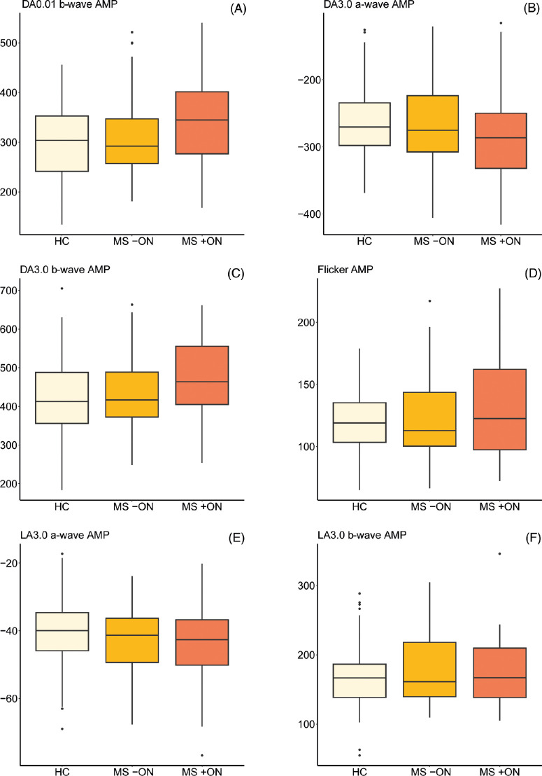
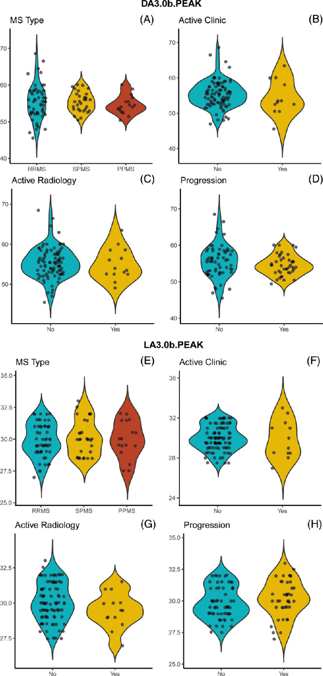
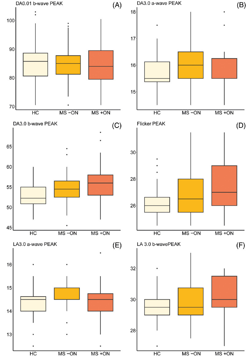
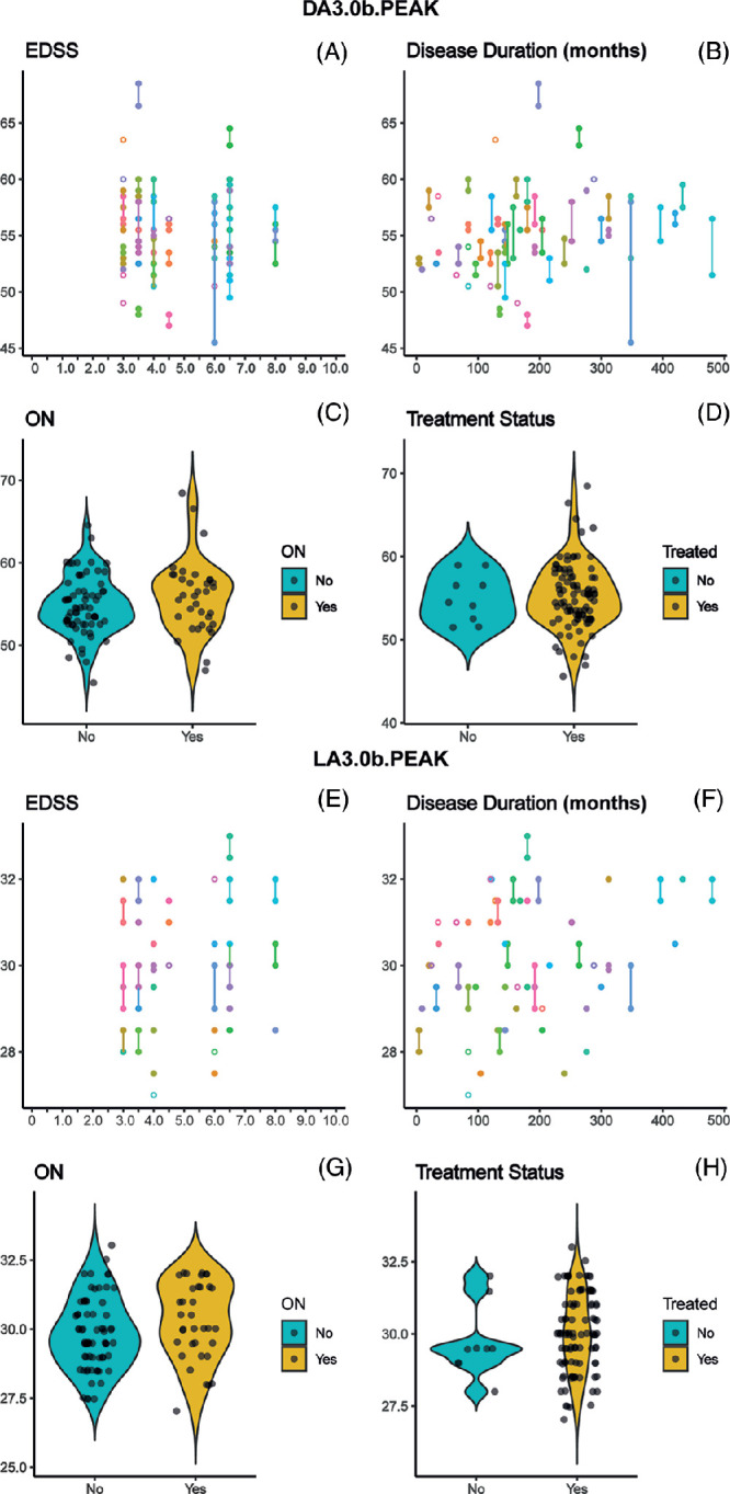
This cross-sectional observational study included pwMS with Expanded Disability Status Scale (EDSS) scores of ≥3.0 and healthy control (HC) participants who underwent ERG, optical coherence tomography, high- and low-contrast visual acuity measurement, and an ophthalmological examination. ERG findings in MS eyes with and without previous optic neuritis (MS +ON; MS -ON) were compared with those in HC eyes. Effects of EDSS, disease duration, ON, and treatment status on selected ERG outcomes were measured. Additional exploratory analyses assessed potential influences of MS phenotype and disease status (clinically active, radiologically active, and disease progression).

RESULTS

Delays to two ERG peak times (dark-adapted 3.0 b-wave; light-adapted flicker) were recorded in MS +ON and MS -ON eyes. No influences of EDSS score, disease duration, previous ON, or treatment status were observed. Exploratory analyses were consistent with no effects of MS phenotype or disease status.

CONCLUSIONS

ERG findings are abnormal in individuals with moderate-severe disability caused by MS; however, these findings are not distinct from those observed earlier in the disease course. Although bipolar dysfunction appears to be common in pwMS throughout the disease course, ERG is likely not useful in monitoring or prognostication of MS.

摘要

目的

多发性硬化症(pwMS)患者的中枢神经系统会出现自身免疫介导的炎症和神经退行性变。目前仍需要一种临床可及、可靠的多发性硬化症神经退行性变的功能标志物。先前的研究已经描述了疾病早期 pwMS 视网膜双极细胞功能的视网膜电图(ERG)衍生测量值的变化。因此,我们研究了 ERG 在疾病进展更严重的个体中的潜在结局测量值。

方法

这项横断面观察性研究纳入了扩展残疾状况量表(EDSS)评分≥3.0 的 pwMS 患者和健康对照(HC)参与者,他们接受了 ERG、光学相干断层扫描、高低对比度视力测量和眼科检查。比较了 MS 眼(有或无先前视神经炎(MS +ON;MS -ON))和 HC 眼的 ERG 结果。测量了 EDSS、疾病持续时间、ON 和治疗状态对选定的 ERG 结果的影响。额外的探索性分析评估了 MS 表型和疾病状态(临床活动、放射学活动和疾病进展)的潜在影响。

结果

在 MS +ON 和 MS -ON 眼中记录到两个 ERG 峰值时间(暗适应 3.0 b 波;明适应闪烁)的延迟。未观察到 EDSS 评分、疾病持续时间、先前的 ON 或治疗状态的影响。探索性分析结果与 MS 表型或疾病状态无影响一致。

结论

在由 MS 引起的中度至重度残疾个体中,ERG 结果异常;然而,这些发现与疾病早期观察到的结果没有明显区别。尽管双极功能障碍似乎在整个疾病过程中在 pwMS 中很常见,但 ERG 不太可能用于监测或预测 MS。

https://cdn.ncbi.nlm.nih.gov/pmc/blobs/a60c/11539974/22f32aa8ad9a/iovs-65-13-2-f004.jpg
https://cdn.ncbi.nlm.nih.gov/pmc/blobs/a60c/11539974/24e9813e9f7b/iovs-65-13-2-f001.jpg
https://cdn.ncbi.nlm.nih.gov/pmc/blobs/a60c/11539974/be1c6c2a3df9/iovs-65-13-2-f002.jpg
https://cdn.ncbi.nlm.nih.gov/pmc/blobs/a60c/11539974/708acbb22e0a/iovs-65-13-2-f003.jpg
https://cdn.ncbi.nlm.nih.gov/pmc/blobs/a60c/11539974/22f32aa8ad9a/iovs-65-13-2-f004.jpg

相似文献

1
Retinal Function in Advanced Multiple Sclerosis.晚期多发性硬化症的视网膜功能。
Invest Ophthalmol Vis Sci. 2024 Nov 4;65(13):2. doi: 10.1167/iovs.65.13.2.
2
A three-year longitudinal study of retinal function and structure in patients with multiple sclerosis.一项针对多发性硬化症患者视网膜功能和结构的三年纵向研究。
Doc Ophthalmol. 2022 Feb;144(1):3-16. doi: 10.1007/s10633-021-09855-7. Epub 2021 Oct 27.
3
Evaluation of the Innermost Retinal Layers and Visual Evoked Potentials in Patients with Multiple Sclerosis.多发性硬化症患者最内层视网膜层及视觉诱发电位的评估
Curr Eye Res. 2016 Oct;41(10):1353-1358. doi: 10.3109/02713683.2015.1119283. Epub 2016 Feb 16.
4
Retinal changes in multiple sclerosis: An optical coherence tomography and angiography study.多发性硬化症的视网膜变化:光学相干断层扫描和血管造影研究。
Rev Neurol (Paris). 2024 Sep;180(7):622-631. doi: 10.1016/j.neurol.2023.11.014. Epub 2024 Mar 7.
5
Outer Retinal Dysfunction in the Absence of Structural Abnormalities in Multiple Sclerosis.多发性硬化症中无结构异常的视网膜外层功能障碍。
Invest Ophthalmol Vis Sci. 2018 Jan 1;59(1):549-560. doi: 10.1167/iovs.17-22821.
6
Analysis of multiple sclerosis patients with electrophysiological and structural tests.对多发性硬化症患者进行电生理和结构测试分析。
Int Ophthalmol. 2017 Jun;37(3):649-653. doi: 10.1007/s10792-016-0324-2. Epub 2016 Aug 18.
7
Evaluation of retinal nerve fiber layer thickness measured by optical coherence tomography in Moroccan patients with multiple sclerosis.光学相干断层扫描测量摩洛哥多发性硬化症患者视网膜神经纤维层厚度的评估
J Fr Ophtalmol. 2015 Jun;38(6):497-503. doi: 10.1016/j.jfo.2014.11.008. Epub 2015 Apr 18.
8
Optical coherence tomography in multiple sclerosis: A Tunisian tertiary center study.多发性硬化症中的光学相干断层扫描:突尼斯三级中心研究。
J Fr Ophtalmol. 2025 Feb;48(2):104371. doi: 10.1016/j.jfo.2024.104371. Epub 2024 Dec 10.
9
Retinal layer segmentation in patients with multiple sclerosis using spectral domain optical coherence tomography.基于谱域光学相干断层扫描的多发性硬化症患者视网膜层分割。
Ophthalmology. 2014 Feb;121(2):573-9. doi: 10.1016/j.ophtha.2013.09.035. Epub 2013 Nov 20.
10
Alterations in the retinal vasculature occur in multiple sclerosis and exhibit novel correlations with disability and visual function measures.视网膜血管的改变发生在多发性硬化症中,并与残疾和视觉功能测量具有新的相关性。
Mult Scler. 2020 Jun;26(7):815-828. doi: 10.1177/1352458519845116. Epub 2019 May 16.

本文引用的文献

1
Multiple sclerosis.多发性硬化症。
Lancet. 2024 Jan 13;403(10422):183-202. doi: 10.1016/S0140-6736(23)01473-3. Epub 2023 Nov 7.
2
The origins of the full-field flash electroretinogram b-wave.全视野闪光视网膜电图b波的起源。
Front Mol Neurosci. 2023 Jul 3;16:1153934. doi: 10.3389/fnmol.2023.1153934. eCollection 2023.
3
Reduced Cone Density Is Associated with Multiple Sclerosis.视锥细胞密度降低与多发性硬化症相关。
Ophthalmol Sci. 2023 Apr 13;3(3):100308. doi: 10.1016/j.xops.2023.100308. eCollection 2023 Sep.
4
ISCEV Standard for full-field clinical electroretinography (2022 update).国际临床电生理学会标准:全视野临床视网膜电流图(2022 更新版)。
Doc Ophthalmol. 2022 Jun;144(3):165-177. doi: 10.1007/s10633-022-09872-0. Epub 2022 May 5.
5
Diagnosis of multiple sclerosis using multifocal ERG data feature fusion.利用多焦视网膜电图数据特征融合诊断多发性硬化症。
Inf Fusion. 2021 Dec;76:157-167. doi: 10.1016/j.inffus.2021.05.006.
6
A three-year longitudinal study of retinal function and structure in patients with multiple sclerosis.一项针对多发性硬化症患者视网膜功能和结构的三年纵向研究。
Doc Ophthalmol. 2022 Feb;144(1):3-16. doi: 10.1007/s10633-021-09855-7. Epub 2021 Oct 27.
7
Negative electroretinograms: genetic and acquired causes, diagnostic approaches and physiological insights.负视网膜电图:遗传和获得性病因、诊断方法和生理见解。
Eye (Lond). 2021 Sep;35(9):2419-2437. doi: 10.1038/s41433-021-01604-z. Epub 2021 Jun 14.
8
A manifesto for reproducible science.可重复科学宣言。
Nat Hum Behav. 2017 Jan 10;1(1):0021. doi: 10.1038/s41562-016-0021.
9
Functional Assessment of Outer and Middle Macular Layers in Multiple Sclerosis.多发性硬化中外层和中层黄斑层的功能评估
J Clin Med. 2020 Nov 22;9(11):3766. doi: 10.3390/jcm9113766.
10
Full-field electroretinography in age-related macular degeneration: an overall retinal response.年龄相关性黄斑变性的全视野视网膜电图:整体视网膜反应。
Acta Ophthalmol. 2021 Mar;99(2):e253-e259. doi: 10.1111/aos.14571. Epub 2020 Aug 24.