• 文献检索
  • 文档翻译
  • 深度研究
  • 学术资讯
  • Suppr Zotero 插件Zotero 插件
  • 邀请有礼
  • 套餐&价格
  • 历史记录
应用&插件
Suppr Zotero 插件Zotero 插件浏览器插件Mac 客户端Windows 客户端微信小程序
定价
高级版会员购买积分包购买API积分包
服务
文献检索文档翻译深度研究API 文档MCP 服务
关于我们
关于 Suppr公司介绍联系我们用户协议隐私条款
关注我们

Suppr 超能文献

核心技术专利:CN118964589B侵权必究
粤ICP备2023148730 号-1Suppr @ 2026

文献检索

告别复杂PubMed语法,用中文像聊天一样搜索,搜遍4000万医学文献。AI智能推荐,让科研检索更轻松。

立即免费搜索

文件翻译

保留排版,准确专业,支持PDF/Word/PPT等文件格式,支持 12+语言互译。

免费翻译文档

深度研究

AI帮你快速写综述,25分钟生成高质量综述,智能提取关键信息,辅助科研写作。

立即免费体验

前列腺癌初始分期和生化复发时采用 Ga PSMA-11 PET 联合 CT 尿路造影方案。

Ga PSMA-11 PET with CT urography protocol in the initial staging and biochemical relapse of prostate cancer.

机构信息

Centre for Molecular Imaging, Department of Cancer Imaging, Peter MacCallum Cancer Centre, 305 Grattan Street, Melbourne, Australia.

Sir Peter MacCallum Department of Oncology, University of Melbourne, 305 Grattan Street, Melbourne, Australia.

出版信息

Cancer Imaging. 2017 Dec 21;17(1):31. doi: 10.1186/s40644-017-0133-5.

DOI:10.1186/s40644-017-0133-5
PMID:29268784
原文链接:https://pmc.ncbi.nlm.nih.gov/articles/PMC5740783/
Abstract

BACKGROUND

Ga-labelled prostate specific membrane antigen (PSMA) ligand PET/CT is a promising modality in primary staging (PS) and biochemical relapse (BCR) of prostate cancer (PC). However, pelvic nodes or local recurrences can be difficult to differentiate from radioactive urine. CT urography (CT-U) is an established method, which allows assessment of urological malignancies. The study presents a novel protocol of Ga-PSMA-11 PET/CT-U in PS and BCR of PC.

METHODS

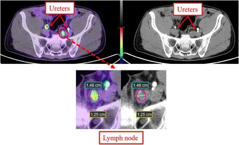
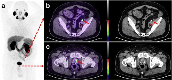
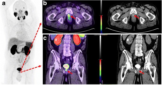
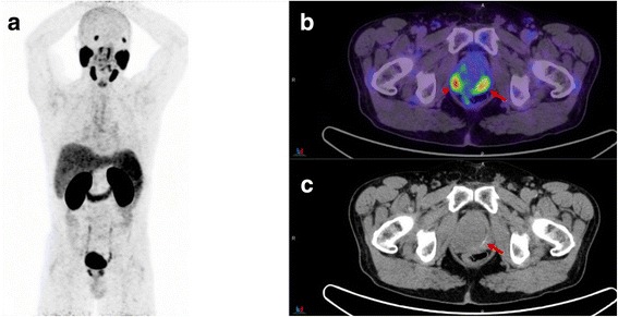
A retrospective review of PSMA PET/CT-U preformed on 57 consecutive patients with prostate cancer. Fifty mL of IV contrast was administered 10 min (range 8-15) before the CT component of a combined PET/CT study, acquired approximately 60 min (range 40-85) after administration of 166 MBq (range 91-246) of Ga-PSMA-11. PET and PET/CT-U were reviewed by two nuclear medicine physicians and CT-U by a radiologist. First, PET images were reviewed independently followed by PET/CT-U images. Foci of activity which could not unequivocally be assessed as disease or urinary activity were recorded. PET/CT-U was considered of potential benefit in final interpretation when the equivocal focal activity in PET images corresponded to opacified ureter, bladder, prostate bed, seminal vesicles, or urethra. Student's T test and Pearson's correlation coefficient was used for assessment of variables including lymph node size and standardized uptake value.

RESULTS

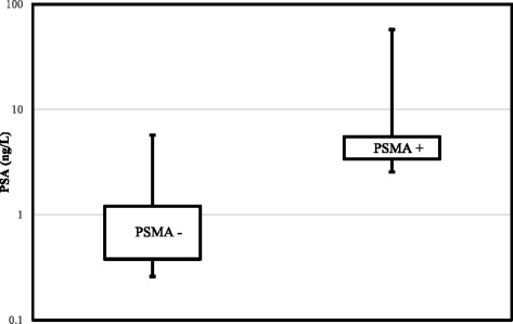
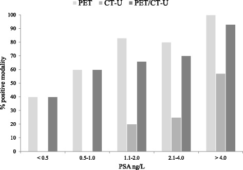
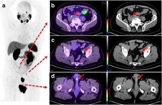
Overall 50 PSMA PET/CT-U studies were performed for BCR and 7 for PS. Median PSA with BCR and PS were 2.0 ± 11.4 ng/ml (0.06-57.3 ng/ml) and 18 ± 35.3 ng/ml (6.8-100 ng/ml), respectively. The median Gleason-score for both groups was 7 (range 6-10). In BCR group, PSMA PET was reported positive in 36 (72%) patients, CT-U in 11(22%) patients and PET/CT-U in 33 (66%) patients. In PS group, PSMA PET detected the primary site in all seven patients, of which one patient with metastatic nodal disease had negative CT finding. Of 40 equivocal foci (27/57 patients) on PET, 11 foci (10/57 patients, 17.5%) were localized to enhanced urine on PET/CT-U, hence considered of potential benefit in interpretation. Of those, 3 foci (3 patients) were solitary sites of activity on PSMA imaging including two local and one nodal site and 4 foci (3 patients) were in different nodal fields.

CONCLUSIONS

PET/CT-U protocol is a practical approach and may assist in interpretation of Ga-PSMA-11 imaging by delineation of the contrast opacified genitourinary system and matching focal PSMA activity with urinary contrast.

摘要

背景

镓标记的前列腺特异性膜抗原(PSMA)配体 PET/CT 是一种有前途的方法,可用于前列腺癌(PC)的原发分期(PS)和生化复发(BCR)。然而,盆腔淋巴结或局部复发可能难以与放射性尿液区分。尿路 CT 造影(CT-U)是一种成熟的方法,可用于评估泌尿系统恶性肿瘤。本研究提出了一种新型的 Ga-PSMA-11 PET/CT-U 在 PS 和 BCR 中的应用方案。

方法

回顾性分析了 57 例连续前列腺癌患者的 Ga-PSMA-11 PET/CT-U 检查结果。在进行 PET/CT 联合研究的 CT 成分前 10 分钟(范围 8-15 分钟)静脉注射 50ml 对比剂,在注射 166MBq(范围 91-246)Ga-PSMA-11 后约 60 分钟(范围 40-85 分钟)进行。两名核医学医师对 PET 和 PET/CT-U 进行了评估,放射科医师对 CT-U 进行了评估。首先,独立评估 PET 图像,然后评估 PET/CT-U 图像。记录无法明确评估为疾病或尿液活动的活动焦点。当 PET 图像中的可疑局灶性活性与显影的输尿管、膀胱、前列腺床、精囊或尿道相对应时,认为 PET/CT-U 对最终解释具有潜在益处。使用学生 t 检验和 Pearson 相关系数评估包括淋巴结大小和标准化摄取值在内的变量。

结果

共有 50 例 BCR 和 7 例 PS 的患者进行了 Ga-PSMA-11 PET/CT-U 检查。BCR 和 PS 组的中位 PSA 分别为 2.0±11.4ng/ml(0.06-57.3ng/ml)和 18±35.3ng/ml(6.8-100ng/ml)。两组的中位 Gleason 评分均为 7(范围 6-10)。在 BCR 组中,36 例(72%)患者的 PSMA PET 报告为阳性,11 例(22%)患者的 CT-U 报告为阳性,33 例(66%)患者的 PET/CT-U 报告为阳性。在 PS 组中,PSMA PET 检测到了所有 7 例患者的原发性疾病,其中 1 例转移性淋巴结疾病患者的 CT 检查为阴性。在 40 个可疑病灶(57 例患者中的 27 个)中,11 个病灶(57 例患者中的 10 个,17.5%)在 PET/CT-U 上定位到增强的尿液,因此认为在解释中具有潜在益处。其中,3 个病灶(3 例患者)为 PSMA 成像中的孤立性活性部位,包括 2 个局部和 1 个淋巴结部位,4 个病灶(3 例患者)位于不同的淋巴结区域。

结论

PET/CT-U 方案是一种实用的方法,通过描绘对比剂显影的泌尿生殖系统并将局灶性 PSMA 活性与尿路对比剂相匹配,可协助 Ga-PSMA-11 成像的解释。

https://cdn.ncbi.nlm.nih.gov/pmc/blobs/52eb/5740783/ee6e84769423/40644_2017_133_Fig7_HTML.jpg
https://cdn.ncbi.nlm.nih.gov/pmc/blobs/52eb/5740783/ec9dd95b6f5d/40644_2017_133_Fig1_HTML.jpg
https://cdn.ncbi.nlm.nih.gov/pmc/blobs/52eb/5740783/add676bf2c67/40644_2017_133_Fig2_HTML.jpg
https://cdn.ncbi.nlm.nih.gov/pmc/blobs/52eb/5740783/0fe70f7d421b/40644_2017_133_Fig3_HTML.jpg
https://cdn.ncbi.nlm.nih.gov/pmc/blobs/52eb/5740783/8691be3fac27/40644_2017_133_Fig4_HTML.jpg
https://cdn.ncbi.nlm.nih.gov/pmc/blobs/52eb/5740783/0cea3c5d771b/40644_2017_133_Fig5_HTML.jpg
https://cdn.ncbi.nlm.nih.gov/pmc/blobs/52eb/5740783/cb2416a363b4/40644_2017_133_Fig6_HTML.jpg
https://cdn.ncbi.nlm.nih.gov/pmc/blobs/52eb/5740783/ee6e84769423/40644_2017_133_Fig7_HTML.jpg

相似文献

1
Ga PSMA-11 PET with CT urography protocol in the initial staging and biochemical relapse of prostate cancer.前列腺癌初始分期和生化复发时采用 Ga PSMA-11 PET 联合 CT 尿路造影方案。
Cancer Imaging. 2017 Dec 21;17(1):31. doi: 10.1186/s40644-017-0133-5.
2
More advantages in detecting bone and soft tissue metastases from prostate cancer using F-PSMA PET/CT.使用F-PSMA PET/CT检测前列腺癌骨和软组织转移方面有更多优势。
Hell J Nucl Med. 2019 Jan-Apr;22(1):6-9. doi: 10.1967/s002449910952. Epub 2019 Mar 7.
3
Multiphasic Ga-PSMA PET/CT in the Detection of Early Recurrence in Prostate Cancer Patients with a PSA Level of Less Than 1 ng/mL: A Prospective Study of 135 Patients.多期 Ga-PSMA PET/CT 在 PSA 水平<1ng/mL 的前列腺癌患者早期复发检测中的应用:一项对 135 例患者的前瞻性研究。
J Nucl Med. 2020 Oct;61(10):1484-1490. doi: 10.2967/jnumed.119.238071. Epub 2020 Feb 14.
4
Early PET imaging with [68]Ga-PSMA-11 increases the detection rate of local recurrence in prostate cancer patients with biochemical recurrence.使用[68]Ga-PSMA-11进行早期PET成像可提高生化复发的前列腺癌患者局部复发的检出率。
Eur J Nucl Med Mol Imaging. 2017 Sep;44(10):1647-1655. doi: 10.1007/s00259-017-3743-z. Epub 2017 Jun 6.
5
Early dynamic imaging in Ga- PSMA-11 PET/CT allows discrimination of urinary bladder activity and prostate cancer lesions.镓-PSMA-11正电子发射断层显像/计算机断层扫描(PET/CT)中的早期动态成像能够区分膀胱活动和前列腺癌病灶。
Eur J Nucl Med Mol Imaging. 2017 May;44(5):765-775. doi: 10.1007/s00259-016-3578-z. Epub 2016 Nov 29.
6
Ga-PSMA PET/CT in patients with recurrent prostate cancer after radical treatment: prospective results in 314 patients.镓-PSMA PET/CT 在根治性治疗后复发前列腺癌患者中的应用:314 例患者的前瞻性结果。
Eur J Nucl Med Mol Imaging. 2018 Nov;45(12):2035-2044. doi: 10.1007/s00259-018-4067-3. Epub 2018 Jun 19.
7
Ga-Labeled Prostate-specific Membrane Antigen Ligand Positron Emission Tomography/Computed Tomography for Prostate Cancer: A Systematic Review and Meta-analysis.Ga 标记前列腺特异性膜抗原配体正电子发射断层扫描/计算机断层扫描在前列腺癌中的应用:系统评价和荟萃分析。
Eur Urol Focus. 2018 Sep;4(5):686-693. doi: 10.1016/j.euf.2016.11.002. Epub 2016 Nov 15.
8
Ga-PSMA PET/CT and Volumetric Morphology of PET-Positive Lymph Nodes Stratified by Tumor Differentiation of Prostate Cancer.基于前列腺癌肿瘤分化分层的Ga-PSMA PET/CT与PET阳性淋巴结的体积形态学
J Nucl Med. 2017 Dec;58(12):1949-1955. doi: 10.2967/jnumed.116.185033. Epub 2017 Jun 21.
9
Ga-PSMA-11 PET/CT in primary staging of prostate cancer: PSA and Gleason score predict the intensity of tracer accumulation in the primary tumour.镓-PSMA-11 PET/CT在前列腺癌初始分期中的应用:前列腺特异抗原(PSA)和 Gleason评分可预测原发肿瘤中示踪剂摄取强度。
Eur J Nucl Med Mol Imaging. 2017 Jun;44(6):941-949. doi: 10.1007/s00259-017-3631-6. Epub 2017 Jan 31.
10
Ga-PSMA-11 PET/CT in recurrent prostate cancer: efficacy in different clinical stages of PSA failure after radical therapy.镓-PSMA-11 PET/CT 在复发性前列腺癌中的应用:根治性治疗后 PSA 失败不同临床阶段的疗效。
Eur J Nucl Med Mol Imaging. 2019 Jan;46(1):31-39. doi: 10.1007/s00259-018-4189-7. Epub 2018 Oct 22.

引用本文的文献

1
Comparative Analysis of Ga-Prostate-Specific Membrane Antigen Positron Emission Tomography/Computed Tomography and Multiparametric Magnetic Resonance Imaging for Gross Tumor Volume Delineation in Radiation Therapy Planning of Prostate Cancer.镓-前列腺特异性膜抗原正电子发射断层扫描/计算机断层扫描与多参数磁共振成像在前列腺癌放射治疗计划中大体肿瘤体积勾画的比较分析
Adv Radiat Oncol. 2025 Jun 9;10(8):101821. doi: 10.1016/j.adro.2025.101821. eCollection 2025 Aug.
2
Distinguishing Physiological Ureter Uptake From an Involved Lymph Node in Staging Prostate-Specific Membrane Antigen (PSMA) Scans: Implications for Radiation Planning.在前列腺特异性膜抗原(PSMA)扫描分期中区分生理性输尿管摄取与受累淋巴结:对放射治疗计划的影响。
Cureus. 2024 Jun 25;16(6):e63105. doi: 10.7759/cureus.63105. eCollection 2024 Jun.
3

本文引用的文献

1
Local recurrence of prostate cancer after radical prostatectomy is at risk to be missed in Ga-PSMA-11-PET of PET/CT and PET/MRI: comparison with mpMRI integrated in simultaneous PET/MRI.前列腺癌根治术后的局部复发在PET/CT和PET/MRI的镓-PSMA-11-PET检查中可能被漏诊:与同时PET/MRI中整合的多参数MRI对比。
Eur J Nucl Med Mol Imaging. 2017 May;44(5):776-787. doi: 10.1007/s00259-016-3594-z. Epub 2016 Dec 17.
2
F-18 labelled PSMA-1007: biodistribution, radiation dosimetry and histopathological validation of tumor lesions in prostate cancer patients.F-18标记的PSMA-1007:前列腺癌患者肿瘤病灶的生物分布、辐射剂量测定及组织病理学验证
Eur J Nucl Med Mol Imaging. 2017 Apr;44(4):678-688. doi: 10.1007/s00259-016-3573-4. Epub 2016 Nov 26.
3
Development and first-in-human study of PSMA-targeted PET tracers with improved pharmacokinetic properties.PSMA 靶向 PET 示踪剂的开发及人体首秀:改善药代动力学特性。
Eur J Nucl Med Mol Imaging. 2024 Jul;51(9):2819-2832. doi: 10.1007/s00259-024-06726-6. Epub 2024 Apr 29.
4
Reducing False-Positives Due to Urinary Stagnation in the Prostatic Urethra on 18 F-DCFPyL PSMA PET/CT With MRI.通过 MRI 降低 18F-DCFPyL PSMA PET/CT 中因前列腺尿道梗阻引起的假阳性。
Clin Nucl Med. 2024 Jul 1;49(7):630-636. doi: 10.1097/RLU.0000000000005220. Epub 2024 Apr 19.
5
Comprehensive literature review of oral and intravenous contrast-enhanced PET/CT: a step forward?口服和静脉注射对比剂增强PET/CT的综合文献综述:是向前迈进了一步吗?
Front Med (Lausanne). 2024 Mar 19;11:1373260. doi: 10.3389/fmed.2024.1373260. eCollection 2024.
6
PSMA PET imaging in the diagnosis and management of prostate cancer.PSMA PET 成像在前列腺癌的诊断和治疗中的应用。
Abdom Radiol (NY). 2023 Dec;48(12):3610-3623. doi: 10.1007/s00261-023-04002-z. Epub 2023 Jul 26.
7
The future of PSMA PET and WB MRI as next-generation imaging tools in prostate cancer.PSMA PET 和 WB MRI 作为前列腺癌下一代成像工具的未来。
Nat Rev Urol. 2022 Aug;19(8):475-493. doi: 10.1038/s41585-022-00618-w. Epub 2022 Jul 4.
8
Appropriate Use Criteria for Prostate-Specific Membrane Antigen PET Imaging.前列腺特异性膜抗原PET成像的合理使用标准
J Nucl Med. 2022 Jan;63(1):59-68. doi: 10.2967/jnumed.121.263262. Epub 2021 Sep 30.
9
Intra-patient comparison of physiologic Ga-PSMA-11 and F-DCFPyL PET/CT uptake in ganglia in prostate cancer patients: a pictorial essay.前列腺癌患者神经节中生理性 Ga-PSMA-11 和 F-DCFPyL PET/CT 摄取的患者内比较:影像学研究。
Cancer Imaging. 2021 Apr 16;21(1):35. doi: 10.1186/s40644-021-00404-8.
10
Combined Intravenous Urogram and 68Ga-PSMA PET/ CT for Improved Staging and Restaging of Prostate Cancer.联合静脉肾盂造影和68Ga-PSMA PET/CT用于改善前列腺癌的分期和再分期
J Clin Imaging Sci. 2020 Oct 24;10:67. doi: 10.25259/JCIS_88_2020. eCollection 2020.
EAU-ESTRO-SIOG Guidelines on Prostate Cancer. Part II: Treatment of Relapsing, Metastatic, and Castration-Resistant Prostate Cancer.EAU-ESTRO-SIOG 前列腺癌诊治指南。第二部分:复发、转移和去势抵抗性前列腺癌的治疗。
Eur Urol. 2017 Apr;71(4):630-642. doi: 10.1016/j.eururo.2016.08.002. Epub 2016 Aug 31.
4
Sensitivity, Specificity, and Predictors of Positive Ga-Prostate-specific Membrane Antigen Positron Emission Tomography in Advanced Prostate Cancer: A Systematic Review and Meta-analysis.前列腺特异性膜抗原正电子发射断层扫描在晚期前列腺癌中的敏感性、特异性和阳性预测值:系统评价和荟萃分析。
Eur Urol. 2016 Dec;70(6):926-937. doi: 10.1016/j.eururo.2016.06.021. Epub 2016 Jun 28.
5
(68)Ga-PSMA ligand PET/CT in patients with prostate cancer: How we review and report.前列腺癌患者的(68)Ga-PSMA配体PET/CT:我们的评估与报告方式
Cancer Imaging. 2016 Jun 8;16(1):14. doi: 10.1186/s40644-016-0072-6.
6
Magnetic resonance imaging for localization of prostate cancer in the setting of biochemical recurrence.用于生化复发情况下前列腺癌定位的磁共振成像
Urol Oncol. 2016 Jul;34(7):303-10. doi: 10.1016/j.urolonc.2016.01.004. Epub 2016 Mar 21.
7
PSMA PET/CT with Glu-urea-Lys-(Ahx)-[⁶⁸Ga(HBED-CC)] versus 3D CT volumetric lymph node assessment in recurrent prostate cancer.用于复发性前列腺癌的PSMA PET/CT与Glu-尿素-Lys-(Ahx)-[⁶⁸Ga(HBED-CC)]对比3D CT容积性淋巴结评估
Eur J Nucl Med Mol Imaging. 2015 Nov;42(12):1794-800. doi: 10.1007/s00259-015-3106-6. Epub 2015 Jul 11.
8
CT Urography for Evaluation of the Ureter.用于评估输尿管的CT尿路造影
Radiographics. 2015 May-Jun;35(3):709-26. doi: 10.1148/rg.2015140209. Epub 2015 Mar 27.
9
Evaluation of PSMA PET/CT imaging using a 68Ga-HBED-CC ligand in patients with prostate cancer and the value of early pelvic imaging.使用68Ga-HBED-CC配体对前列腺癌患者进行PSMA PET/CT成像评估及早期盆腔成像的价值
Nucl Med Commun. 2015 Jun;36(6):582-7. doi: 10.1097/MNM.0000000000000290.
10
Cancer statistics, 2014.癌症统计数据,2014 年。
CA Cancer J Clin. 2014 Jan-Feb;64(1):9-29. doi: 10.3322/caac.21208. Epub 2014 Jan 7.