Wang Liang, Tang Dalin, Maehara Akiko, Wu Zheyang, Yang Chun, Muccigrosso David, Zheng Jie, Bach Richard, Billiar Kristen L, Mintz Gary S
School of Biological Science and Medical Engineering, Southeast University, Nanjing, China; Mathematical Sciences Department, Worcester Polytechnic Institute, MA, USA.
School of Biological Science and Medical Engineering, Southeast University, Nanjing, China; Mathematical Sciences Department, Worcester Polytechnic Institute, MA, USA.
J Biomech. 2018 Feb 8;68:43-50. doi: 10.1016/j.jbiomech.2017.12.007. Epub 2017 Dec 15.
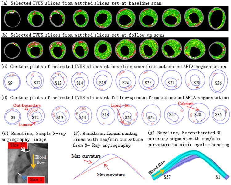
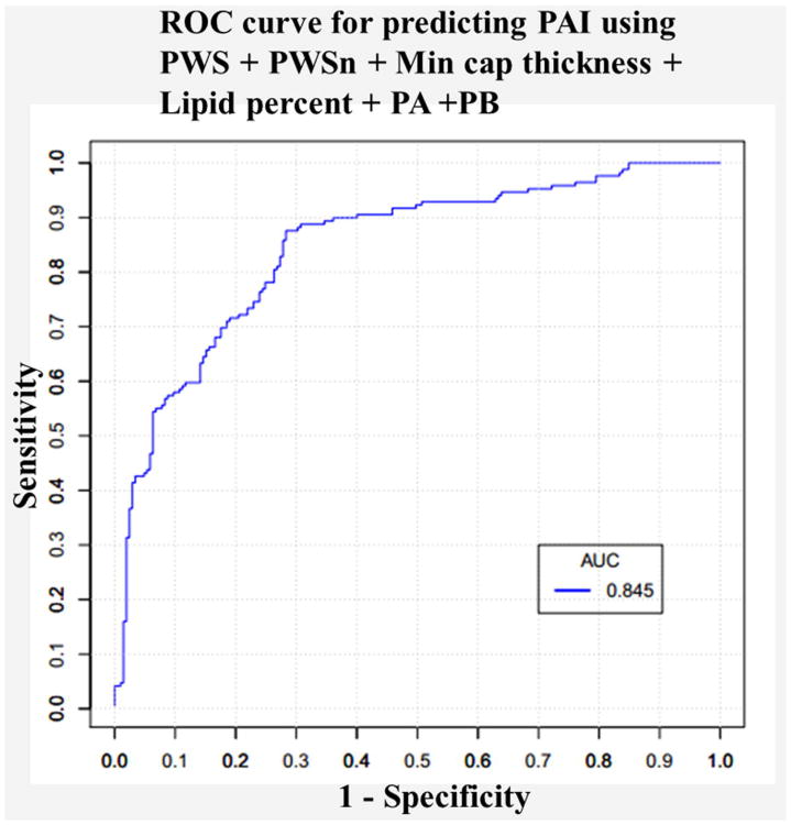
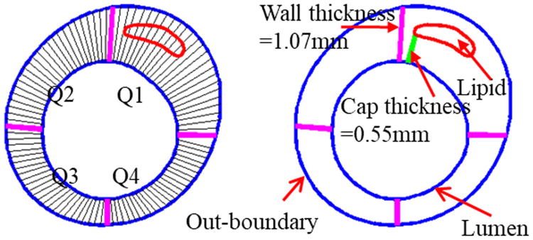
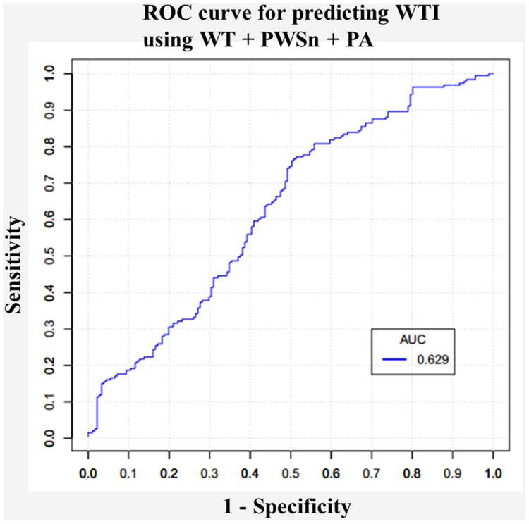
Plaque morphology and biomechanics are believed to be closely associated with plaque progression. In this paper, we test the hypothesis that integrating morphological and biomechanical risk factors would result in better predictive power for plaque progression prediction. A sample size of 374 intravascular ultrasound (IVUS) slices was obtained from 9 patients with IVUS follow-up data. 3D fluid-structure interaction models were constructed to obtain both structural stress/strain and fluid biomechanical conditions. Data for eight morphological and biomechanical risk factors were extracted for each slice. Plaque area increase (PAI) and wall thickness increase (WTI) were chosen as two measures for plaque progression. Progression measure and risk factors were fed to generalized linear mixed models and linear mixed-effect models to perform prediction and correlation analysis, respectively. All combinations of eight risk factors were exhausted to identify the optimal predictor(s) with highest prediction accuracy defined as sum of sensitivity and specificity. When using a single risk factor, plaque wall stress (PWS) at baseline was the best predictor for plaque progression (PAI and WTI). The optimal predictor among all possible combinations for PAI was PWS + PWSn + Lipid percent + Min cap thickness + Plaque Area (PA) + Plaque Burden (PB) (prediction accuracy = 1.5928) while Wall Thickness (WT) + Plaque Wall Strain (PWSn) + Plaque Area (PA) was the best for WTI (1.2589). This indicated that PAI was a more predictable measure than WTI. The combination including both morphological and biomechanical parameters had improved prediction accuracy, compared to predictions using only morphological features.
斑块形态学和生物力学被认为与斑块进展密切相关。在本文中,我们检验了这样一个假设,即整合形态学和生物力学风险因素将对斑块进展预测产生更好的预测能力。从9例有血管内超声(IVUS)随访数据的患者中获取了374个血管内超声切片样本。构建了三维流固相互作用模型,以获得结构应力/应变和流体生物力学条件。为每个切片提取了八个形态学和生物力学风险因素的数据。选择斑块面积增加(PAI)和管壁厚度增加(WTI)作为斑块进展的两个指标。将进展指标和风险因素分别输入广义线性混合模型和线性混合效应模型进行预测和相关性分析。穷尽八个风险因素的所有组合,以确定预测准确性最高的最佳预测指标,预测准确性定义为敏感性和特异性之和。当使用单一风险因素时,基线时的斑块壁应力(PWS)是斑块进展(PAI和WTI)的最佳预测指标。PAI所有可能组合中的最佳预测指标是PWS + PWSn + 脂质百分比 + 最小帽厚度 + 斑块面积(PA)+ 斑块负荷(PB)(预测准确性 = 1.5928),而管壁厚度(WT)+ 斑块壁应变(PWSn)+ 斑块面积(PA)对WTI是最佳的(1.2589)。这表明PAI比WTI是更可预测的指标。与仅使用形态学特征的预测相比,包括形态学和生物力学参数的组合提高了预测准确性。