• 文献检索
  • 文档翻译
  • 深度研究
  • 学术资讯
  • Suppr Zotero 插件Zotero 插件
  • 邀请有礼
  • 套餐&价格
  • 历史记录
应用&插件
Suppr Zotero 插件Zotero 插件浏览器插件Mac 客户端Windows 客户端微信小程序
定价
高级版会员购买积分包购买API积分包
服务
文献检索文档翻译深度研究API 文档MCP 服务
关于我们
关于 Suppr公司介绍联系我们用户协议隐私条款
关注我们

Suppr 超能文献

核心技术专利:CN118964589B侵权必究
粤ICP备2023148730 号-1Suppr @ 2026

文献检索

告别复杂PubMed语法,用中文像聊天一样搜索,搜遍4000万医学文献。AI智能推荐,让科研检索更轻松。

立即免费搜索

文件翻译

保留排版,准确专业,支持PDF/Word/PPT等文件格式,支持 12+语言互译。

免费翻译文档

深度研究

AI帮你快速写综述,25分钟生成高质量综述,智能提取关键信息,辅助科研写作。

立即免费体验

全心脏发育过程中的细胞精度光学标测。

Cell-accurate optical mapping across the entire developing heart.

机构信息

Max Planck Institute of Molecular Cell Biology and Genetics, Dresden, Germany.

Harvard Medical School, Boston, United States.

出版信息

Elife. 2017 Dec 29;6:e28307. doi: 10.7554/eLife.28307.

DOI:10.7554/eLife.28307
PMID:29286002
原文链接:https://pmc.ncbi.nlm.nih.gov/articles/PMC5747520/
Abstract

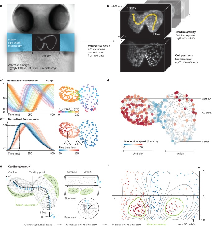
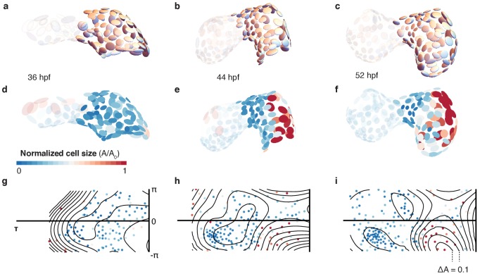
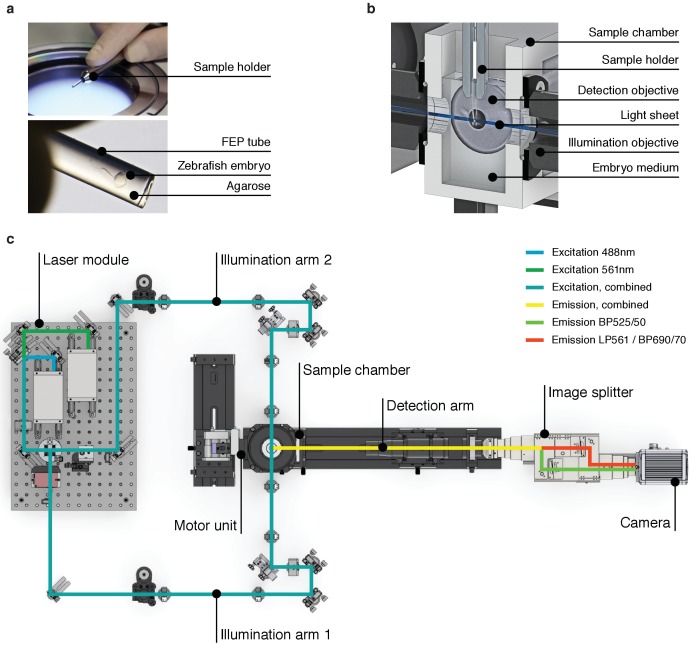
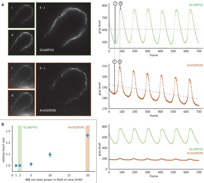
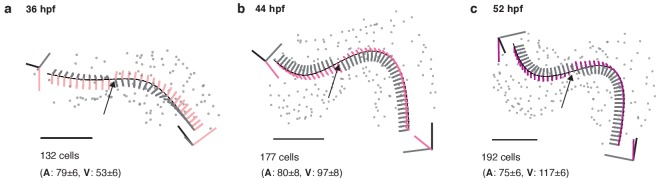
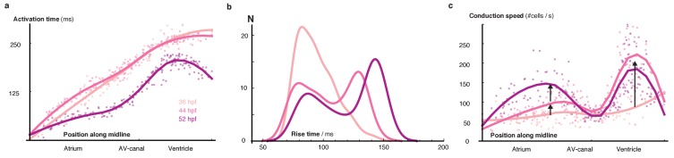
Organogenesis depends on orchestrated interactions between individual cells and morphogenetically relevant cues at the tissue level. This is true for the heart, whose function critically relies on well-ordered communication between neighboring cells, which is established and fine-tuned during embryonic development. For an integrated understanding of the development of structure and function, we need to move from isolated snap-shot observations of either microscopic or macroscopic parameters to simultaneous and, ideally continuous, cell-to-organ scale imaging. We introduce cell-accurate three-dimensional Ca-mapping of all cells in the entire electro-mechanically uncoupled heart during the looping stage of live embryonic zebrafish, using high-speed light sheet microscopy and tailored image processing and analysis. We show how myocardial region-specific heterogeneity in cell function emerges during early development and how structural patterning goes hand-in-hand with functional maturation of the entire heart. Our method opens the way to systematic, scale-bridging, studies of vertebrate organogenesis by cell-accurate structure-function mapping across entire organs.

摘要

器官发生依赖于个体细胞之间的协调相互作用和组织水平上与形态发生相关的线索。心脏就是如此,其功能严重依赖于相邻细胞之间有序的通讯,这种通讯在胚胎发育过程中建立并精细调整。为了综合理解结构和功能的发育,我们需要将对微观或宏观参数的孤立快照观察转变为同时进行的、理想情况下是连续的、从细胞到器官尺度的成像。我们使用高速光片显微镜和定制的图像处理和分析,在活体斑马鱼胚胎的心脏环化阶段,对整个电机械解耦心脏中的所有细胞进行精确到细胞的三维 Ca 映射。我们展示了在早期发育过程中,心肌区域特异性细胞功能异质性是如何出现的,以及结构模式是如何与整个心脏的功能成熟齐头并进的。我们的方法为通过跨越整个器官的精确到细胞的结构-功能映射对脊椎动物器官发生进行系统的、跨尺度的研究开辟了道路。

https://cdn.ncbi.nlm.nih.gov/pmc/blobs/dc88/5747520/4fe8961e52f4/elife-28307-fig2-figsupp3.jpg
https://cdn.ncbi.nlm.nih.gov/pmc/blobs/dc88/5747520/d6780b662f7b/elife-28307-fig1.jpg
https://cdn.ncbi.nlm.nih.gov/pmc/blobs/dc88/5747520/b87542e1f27f/elife-28307-fig1-figsupp1.jpg
https://cdn.ncbi.nlm.nih.gov/pmc/blobs/dc88/5747520/c769616da2fb/elife-28307-fig1-figsupp2.jpg
https://cdn.ncbi.nlm.nih.gov/pmc/blobs/dc88/5747520/370fda9aca96/elife-28307-fig1-figsupp3.jpg
https://cdn.ncbi.nlm.nih.gov/pmc/blobs/dc88/5747520/492b0dae1f4b/elife-28307-fig1-figsupp4.jpg
https://cdn.ncbi.nlm.nih.gov/pmc/blobs/dc88/5747520/51d04ff245b5/elife-28307-fig1-figsupp5.jpg
https://cdn.ncbi.nlm.nih.gov/pmc/blobs/dc88/5747520/f9514e578a31/elife-28307-fig1-figsupp6.jpg
https://cdn.ncbi.nlm.nih.gov/pmc/blobs/dc88/5747520/a678bf8c6982/elife-28307-fig1-figsupp7.jpg
https://cdn.ncbi.nlm.nih.gov/pmc/blobs/dc88/5747520/e1c647c84a67/elife-28307-fig1-figsupp8.jpg
https://cdn.ncbi.nlm.nih.gov/pmc/blobs/dc88/5747520/4e00b15ed858/elife-28307-fig1-figsupp9.jpg
https://cdn.ncbi.nlm.nih.gov/pmc/blobs/dc88/5747520/c704f5e16100/elife-28307-fig2.jpg
https://cdn.ncbi.nlm.nih.gov/pmc/blobs/dc88/5747520/de63ff093295/elife-28307-fig2-figsupp1.jpg
https://cdn.ncbi.nlm.nih.gov/pmc/blobs/dc88/5747520/1eff958593f3/elife-28307-fig2-figsupp2.jpg
https://cdn.ncbi.nlm.nih.gov/pmc/blobs/dc88/5747520/4fe8961e52f4/elife-28307-fig2-figsupp3.jpg

相似文献

1
Cell-accurate optical mapping across the entire developing heart.全心脏发育过程中的细胞精度光学标测。
Elife. 2017 Dec 29;6:e28307. doi: 10.7554/eLife.28307.
2
In vivo imaging of cardiac development and function in zebrafish using light sheet microscopy.利用光片显微镜对斑马鱼心脏发育和功能进行体内成像。
Swiss Med Wkly. 2015 Dec 23;145:w14227. doi: 10.4414/smw.2015.14227. eCollection 2015.
3
Multiscale light-sheet for rapid imaging of cardiopulmonary system.多尺度光片用于心肺系统的快速成像。
JCI Insight. 2018 Aug 23;3(16). doi: 10.1172/jci.insight.121396.
4
Three-dimensional correction of conduction velocity in the embryonic heart using integrated optical mapping and optical coherence tomography.利用集成光学映射和光学相干断层扫描对胚胎心脏传导速度进行三维校正。
J Biomed Opt. 2014;19(7):76004. doi: 10.1117/1.JBO.19.7.076004.
5
Analysis of 4D myocardial wall motion during early stages of chick heart development.鸡胚心脏发育早期4D心肌壁运动分析
Methods Mol Biol. 2015;1299:191-212. doi: 10.1007/978-1-4939-2572-8_16.
6
Morphogenetic control of zebrafish cardiac looping by Bmp signaling.Bmp 信号对斑马鱼心脏环化的形态发生控制。
Development. 2019 Nov 14;146(22):dev180091. doi: 10.1242/dev.180091.
7
Multi-scale imaging and analysis identify pan-embryo cell dynamics of germlayer formation in zebrafish.多尺度成像和分析鉴定斑马鱼胚层形成中的泛胚胎细胞动力学。
Nat Commun. 2019 Dec 17;10(1):5753. doi: 10.1038/s41467-019-13625-0.
8
Imaging the beating heart in the mouse using intravital microscopy techniques.使用活体显微镜技术对小鼠跳动的心脏进行成像。
Nat Protoc. 2015 Nov;10(11):1802-19. doi: 10.1038/nprot.2015.119. Epub 2015 Oct 22.
9
Rapid three-dimensional imaging and analysis of the beating embryonic heart reveals functional changes during development.对跳动的胚胎心脏进行快速三维成像和分析可揭示发育过程中的功能变化。
Dev Dyn. 2006 Nov;235(11):2940-8. doi: 10.1002/dvdy.20926.
10
A combined light sheet fluorescence and differential interference contrast microscope for live imaging of multicellular specimens.一种用于多细胞样本活体成像的联合光片荧光和微分干涉对比显微镜。
J Microsc. 2015 May;258(2):105-12. doi: 10.1111/jmi.12220. Epub 2015 Jan 22.

引用本文的文献

1
Towards advanced regenerative therapeutics to tackle cardio-cerebrovascular diseases.迈向治疗心脑血管疾病的先进再生疗法。
Am Heart J Plus. 2025 Mar 1;53:100520. doi: 10.1016/j.ahjo.2025.100520. eCollection 2025 May.
2
Tissue stiffness mapping by light sheet elastography.通过光片弹性成像进行组织硬度映射
Sci Adv. 2025 Mar 14;11(11):eadt7274. doi: 10.1126/sciadv.adt7274.
3
Regionalized regulation of actomyosin organization influences cardiomyocyte cell shape changes during chamber curvature formation.肌动球蛋白组织的区域化调控影响心室曲率形成过程中心肌细胞的形态变化。

本文引用的文献

1
Axial tubule junctions control rapid calcium signaling in atria.轴突小管连接调控心房中的快速钙信号传导。
J Clin Invest. 2016 Oct 3;126(10):3999-4015. doi: 10.1172/JCI88241. Epub 2016 Sep 19.
2
Chamber identity programs drive early functional partitioning of the heart.腔室识别程序驱动心脏的早期功能分区。
Nat Commun. 2015 Aug 26;6:8146. doi: 10.1038/ncomms9146.
3
Dynamic structure and protein expression of the live embryonic heart captured by 2-photon light sheet microscopy and retrospective registration.通过双光子光片显微镜和回顾性配准捕获的活胚胎心脏的动态结构和蛋白质表达。
bioRxiv. 2025 Jan 8:2025.01.07.631779. doi: 10.1101/2025.01.07.631779.
4
Heart-on-a-chip systems with tissue-specific functionalities for physiological, pathological, and pharmacological studies.具有组织特异性功能的芯片上心脏系统,用于生理、病理和药理研究。
Mater Today Bio. 2023 Dec 20;24:100914. doi: 10.1016/j.mtbio.2023.100914. eCollection 2024 Feb.
5
Topological packing statistics of living and nonliving matter.生物和非生物的拓扑堆积统计。
Sci Adv. 2023 Sep 8;9(36):eadg1261. doi: 10.1126/sciadv.adg1261. Epub 2023 Sep 6.
6
4D Light-sheet imaging and interactive analysis of cardiac contractility in zebrafish larvae.斑马鱼幼体心脏收缩性的4D光片成像与交互式分析
APL Bioeng. 2023 Jun 15;7(2):026112. doi: 10.1063/5.0153214. eCollection 2023 Jun.
7
Compact, high-speed multi-directional selective plane illumination microscopy.紧凑型高速多向选择性平面照明显微镜。
Biomed Opt Express. 2023 Mar 7;14(4):1445-1459. doi: 10.1364/BOE.476217. eCollection 2023 Apr 1.
8
KHz-rate volumetric voltage imaging of the whole Zebrafish heart.斑马鱼整个心脏的千赫兹速率容积电压成像
Biophys Rep (N Y). 2022 Feb 3;2(1):100046. doi: 10.1016/j.bpr.2022.100046. eCollection 2022 Mar 9.
9
Real-Time Optical Mapping of Contracting Cardiac Tissues With GPU-Accelerated Numerical Motion Tracking.利用GPU加速数值运动跟踪对收缩期心脏组织进行实时光学映射。
Front Cardiovasc Med. 2022 May 24;9:787627. doi: 10.3389/fcvm.2022.787627. eCollection 2022.
10
Simultaneous imaging of calcium and contraction in the beating heart of zebrafish larvae.斑马鱼幼鱼心跳过程中钙离子和收缩的同步成像。
Theranostics. 2022 Jan 1;12(3):1012-1029. doi: 10.7150/thno.64734. eCollection 2022.
Biomed Opt Express. 2015 May 11;6(6):2056-66. doi: 10.1364/BOE.6.002056. eCollection 2015 Jun 1.
4
Swept confocally-aligned planar excitation (SCAPE) microscopy for high speed volumetric imaging of behaving organisms.用于行为生物体高速体积成像的扫描共焦对齐平面激发(SCAPE)显微镜。
Nat Photonics. 2015 Feb;9(2):113-119. doi: 10.1038/nphoton.2014.323.
5
Simultaneous mapping of membrane voltage and calcium in zebrafish heart in vivo reveals chamber-specific developmental transitions in ionic currents.在体斑马鱼心脏中膜电压和钙的同步测绘揭示了离子电流中特定腔室的发育转变。
Front Physiol. 2014 Sep 11;5:344. doi: 10.3389/fphys.2014.00344. eCollection 2014.
6
High-resolution reconstruction of the beating zebrafish heart.高分辨率重建斑马鱼跳动的心脏。
Nat Methods. 2014 Sep;11(9):919-22. doi: 10.1038/nmeth.3037. Epub 2014 Jul 20.
7
The zebrafish model system in cardiovascular research: A tiny fish with mighty prospects.心血管研究中的斑马鱼模型系统:前景广阔的小鱼。
Glob Cardiol Sci Pract. 2013 Nov 1;2013(1):9-28. doi: 10.5339/gcsp.2013.4. eCollection 2013.
8
Rapid 3D light-sheet microscopy with a tunable lens.采用可调谐透镜的快速三维光片显微镜。
Opt Express. 2013 Sep 9;21(18):21010-26. doi: 10.1364/OE.21.021010.
9
tal1 Regulates the formation of intercellular junctions and the maintenance of identity in the endocardium.tal1 调控心内膜细胞连接的形成和细胞特性的维持。
Dev Biol. 2013 Nov 15;383(2):214-26. doi: 10.1016/j.ydbio.2013.09.019. Epub 2013 Sep 25.
10
Identification and functional characterization of cardiac pacemaker cells in zebrafish.鉴定和功能表征斑马鱼的心脏起搏细胞。
PLoS One. 2012;7(10):e47644. doi: 10.1371/journal.pone.0047644. Epub 2012 Oct 16.