Clinical Cell Biology, Department of Pathology, Odense University Hospital, 5000 Odense C, Denmark.
Department of Clinical Research, University of Southern Denmark, 5230 Odense M, Denmark.
Int J Mol Sci. 2020 Oct 19;21(20):7717. doi: 10.3390/ijms21207717.
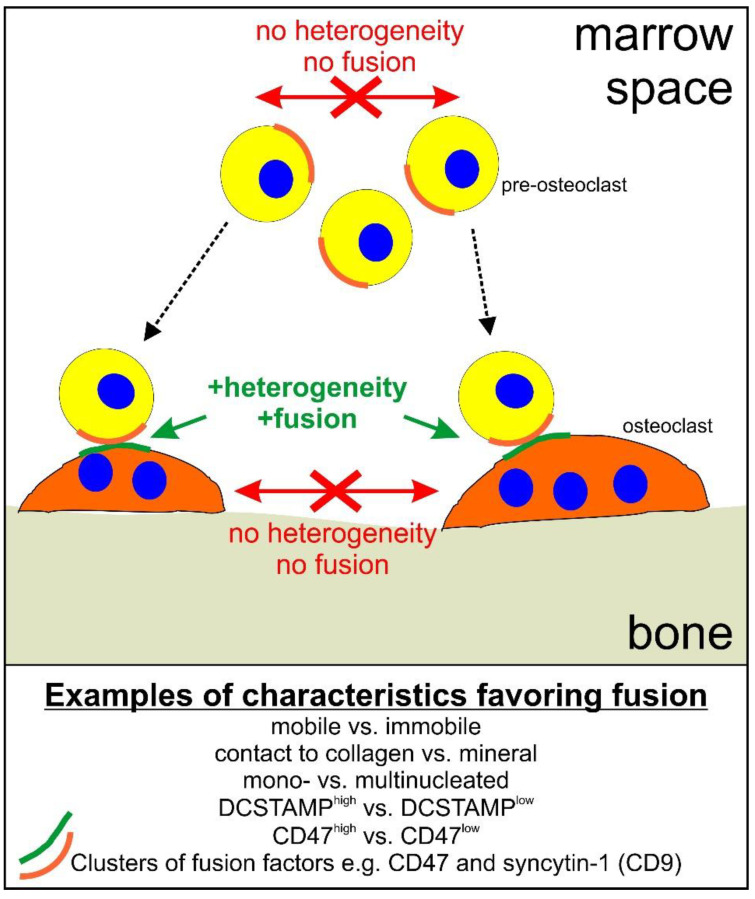
Classically, osteoclast fusion consists of four basic steps: (1) attraction/migration, (2) recognition, (3) cell-cell adhesion, and (4) membrane fusion. In theory, this sounds like a straightforward simple linear process. However, it is not. Osteoclast fusion has to take place in a well-coordinated manner-something that is not simple. In vivo, the complex regulation of osteoclast formation takes place within the bone marrow-in time and space. The present review will focus on considering osteoclast fusion in the context of physiology and pathology. Special attention is given to: (1) regulation of osteoclast fusion in vivo, (2) heterogeneity of osteoclast fusion partners, (3) regulation of multi-nucleation, (4) implications for physiology and pathology, and (5) implications for drug sensitivity and side effects. The review will emphasize that more attention should be given to the human in vivo reality when interpreting the impact of in vitro and animal studies. This should be done in order to improve our understanding of human physiology and pathology, as well as to improve anti-resorptive treatment and reduce side effects.
经典理论认为,破骨细胞融合包含四个基本步骤:(1)吸引/迁移;(2)识别;(3)细胞间黏附;(4)细胞膜融合。理论上,这似乎是一个简单的线性过程。但事实并非如此。破骨细胞融合必须以协调的方式发生——这并不简单。在体内,破骨细胞的形成受到复杂的调控,发生在时间和空间上。本综述将重点讨论生理学和病理学背景下的破骨细胞融合。特别关注以下几点:(1)体内破骨细胞融合的调控;(2)破骨细胞融合伙伴的异质性;(3)多核形成的调控;(4)对生理学和病理学的影响;(5)对药物敏感性和副作用的影响。综述强调,在解释体外和动物研究的影响时,应更加关注人类体内的实际情况。这样做是为了增进我们对人类生理学和病理学的理解,改善抗吸收治疗效果并减少副作用。