• 文献检索
  • 文档翻译
  • 深度研究
  • 学术资讯
  • Suppr Zotero 插件Zotero 插件
  • 邀请有礼
  • 套餐&价格
  • 历史记录
应用&插件
Suppr Zotero 插件Zotero 插件浏览器插件Mac 客户端Windows 客户端微信小程序
定价
高级版会员购买积分包购买API积分包
服务
文献检索文档翻译深度研究API 文档MCP 服务
关于我们
关于 Suppr公司介绍联系我们用户协议隐私条款
关注我们

Suppr 超能文献

核心技术专利:CN118964589B侵权必究
粤ICP备2023148730 号-1Suppr @ 2026

文献检索

告别复杂PubMed语法,用中文像聊天一样搜索,搜遍4000万医学文献。AI智能推荐,让科研检索更轻松。

立即免费搜索

文件翻译

保留排版,准确专业,支持PDF/Word/PPT等文件格式,支持 12+语言互译。

免费翻译文档

深度研究

AI帮你快速写综述,25分钟生成高质量综述,智能提取关键信息,辅助科研写作。

立即免费体验

3特斯拉高精度计算机扩散加权成像诊断肝细胞癌的初步结果

Preliminary Results of High-Precision Computed Diffusion Weighted Imaging for the Diagnosis of Hepatocellular Carcinoma at 3 Tesla.

作者信息

Akagi Motonori, Nakamura Yuko, Higaki Toru, Matsubara Yoshiko, Terada Hiroaki, Honda Yukiko, Tatsugami Fuminari, Baba Yasutaka, Iida Makoto, Awai Kazuo

机构信息

From the Diagnostic Radiology, Hiroshima University, Hiroshima, Japan.

出版信息

J Comput Assist Tomogr. 2018 May/Jun;42(3):373-379. doi: 10.1097/RCT.0000000000000702.

DOI:10.1097/RCT.0000000000000702
PMID:29287019
原文链接:https://pmc.ncbi.nlm.nih.gov/articles/PMC5976220/
Abstract

OBJECTIVE

To compare the utility of high-precision computed diffusion-weighted imaging (hc-DWI) and conventional computed DWI (cc-DWI) for the diagnosis of hepatocellular carcinoma (HCC) at 3 T.

METHODS

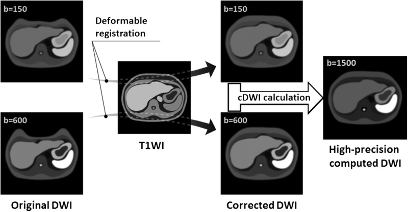
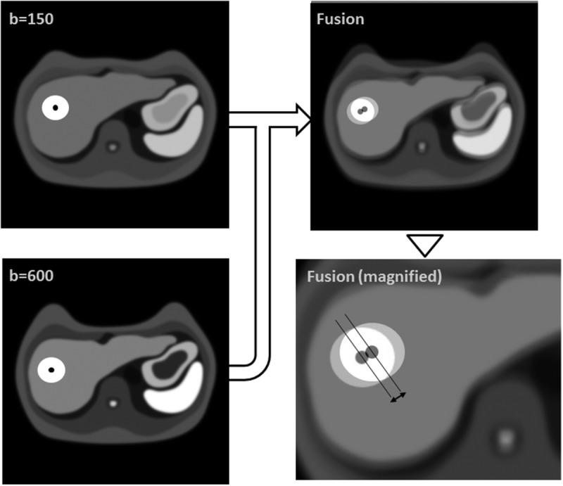
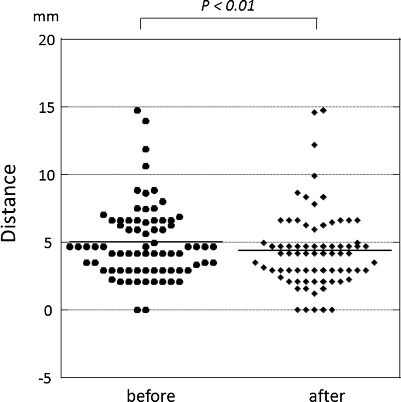
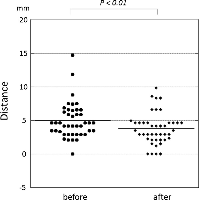
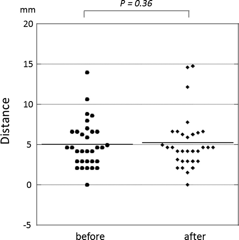
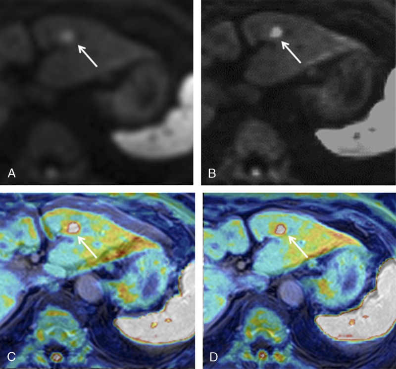
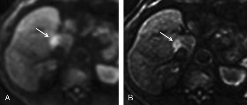
We subjected 75 HCC patients to DWI (b-value 150 and 600 s/mm). To generate hc-DWI we applied non-rigid image registration to avoid the mis-registration of images obtained with different b-values. We defined c-DWI with a b-value of 1500 s/mm using DWI with b-value 150 and 600 s/mm as cc-DWI, and c-DWI with b-value 1500 s/mm using registered DWI with b-value 150 and 600 s/mm as hc-DWI. A radiologist recorded the contrast ratio (CR) between HCC and the surrounding hepatic parenchyma.

RESULTS

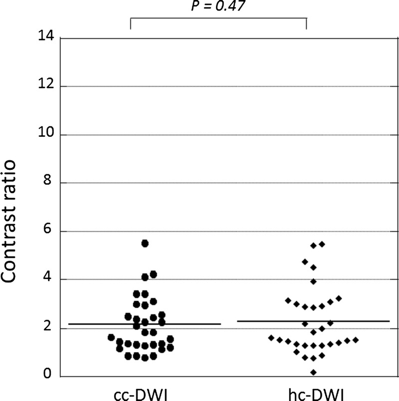
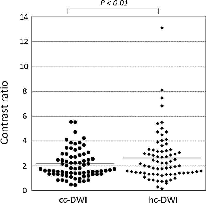
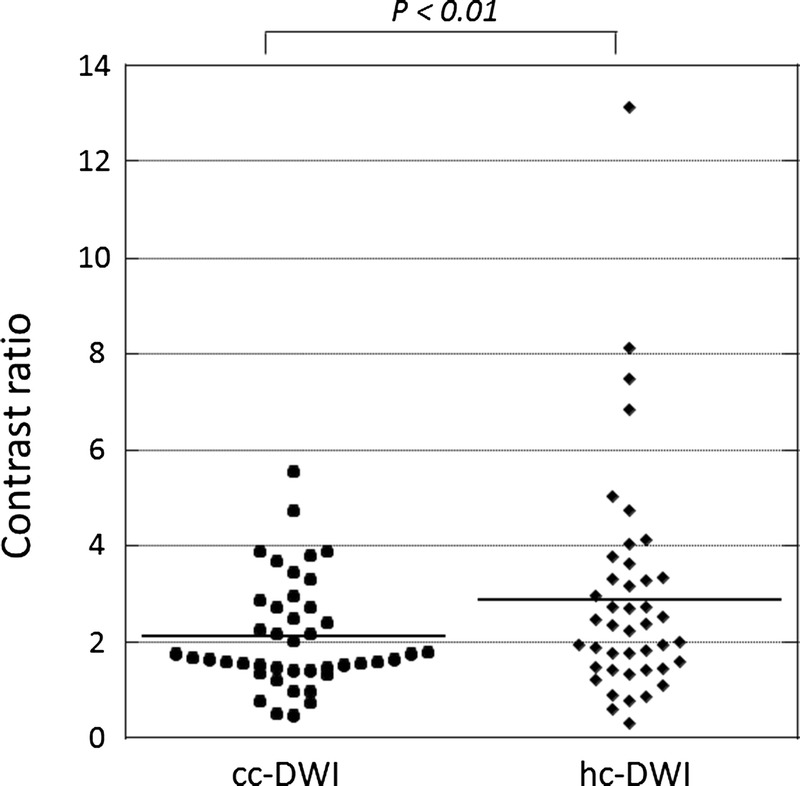
The CR for HCC was significantly higher on hc- than cc-DWIs (median 2.0 vs. 1.8, P < 0.01).

CONCLUSION

The CR of HCC can be improved with image registration, indicating that hc-DWI is more useful than cc-DWI for the diagnosis of HCC.

摘要

目的

比较高精度计算机扩散加权成像(hc-DWI)和传统计算机扩散加权成像(cc-DWI)在3T场强下对肝细胞癌(HCC)的诊断效用。

方法

我们对75例HCC患者进行了扩散加权成像(b值为150和600 s/mm²)。为生成hc-DWI,我们应用了非刚性图像配准以避免不同b值获得的图像配准错误。我们将使用b值为150和600 s/mm²的扩散加权成像定义为b值为1500 s/mm²的c-DWI作为cc-DWI,将使用配准后的b值为150和600 s/mm²的扩散加权成像定义为b值为1500 s/mm²的c-DWI作为hc-DWI。一名放射科医生记录了HCC与周围肝实质之间的对比率(CR)。

结果

hc-DWI上HCC的CR显著高于cc-DWI(中位数2.0对1.8,P < 0.01)。

结论

图像配准可提高HCC的CR,表明hc-DWI在HCC诊断中比cc-DWI更有用。

https://cdn.ncbi.nlm.nih.gov/pmc/blobs/6d95/5976220/7f572c645d78/rct-42-373-g010.jpg
https://cdn.ncbi.nlm.nih.gov/pmc/blobs/6d95/5976220/c3e76106d9a9/rct-42-373-g001.jpg
https://cdn.ncbi.nlm.nih.gov/pmc/blobs/6d95/5976220/8e39e60eec7e/rct-42-373-g002.jpg
https://cdn.ncbi.nlm.nih.gov/pmc/blobs/6d95/5976220/1bdd666ae5e9/rct-42-373-g003.jpg
https://cdn.ncbi.nlm.nih.gov/pmc/blobs/6d95/5976220/82cf8d95eeff/rct-42-373-g004.jpg
https://cdn.ncbi.nlm.nih.gov/pmc/blobs/6d95/5976220/cb4f262e100c/rct-42-373-g005.jpg
https://cdn.ncbi.nlm.nih.gov/pmc/blobs/6d95/5976220/002bcb4c6ae5/rct-42-373-g006.jpg
https://cdn.ncbi.nlm.nih.gov/pmc/blobs/6d95/5976220/f3a39df3c18f/rct-42-373-g007.jpg
https://cdn.ncbi.nlm.nih.gov/pmc/blobs/6d95/5976220/40eb5adb3642/rct-42-373-g008.jpg
https://cdn.ncbi.nlm.nih.gov/pmc/blobs/6d95/5976220/4dbc1bd6bbf3/rct-42-373-g009.jpg
https://cdn.ncbi.nlm.nih.gov/pmc/blobs/6d95/5976220/7f572c645d78/rct-42-373-g010.jpg

相似文献

1
Preliminary Results of High-Precision Computed Diffusion Weighted Imaging for the Diagnosis of Hepatocellular Carcinoma at 3 Tesla.3特斯拉高精度计算机扩散加权成像诊断肝细胞癌的初步结果
J Comput Assist Tomogr. 2018 May/Jun;42(3):373-379. doi: 10.1097/RCT.0000000000000702.
2
Hepatocellular carcinoma: Can LI-RADS v2017 with gadoxetic-acid enhancement magnetic resonance and diffusion-weighted imaging improve diagnostic accuracy?肝细胞癌:钆塞酸增强磁共振和弥散加权成像的 LI-RADS v2017 能否提高诊断准确性?
World J Gastroenterol. 2019 Feb 7;25(5):622-631. doi: 10.3748/wjg.v25.i5.622.
3
Diffusion kurtosis imaging of the liver at 3 Tesla: in vivo comparison to standard diffusion-weighted imaging.3特斯拉下肝脏的扩散峰度成像:与标准扩散加权成像的体内比较
Acta Radiol. 2018 Jan;59(1):18-25. doi: 10.1177/0284185117706608. Epub 2017 Apr 28.
4
Diffusion-weighted imaging of the liver in patients with chronic liver disease: comparison of monopolar and bipolar diffusion gradients for image quality and lesion detection.慢性肝病患者肝脏的弥散加权成像:单极和双极弥散梯度在图像质量和病灶检测方面的比较。
AJR Am J Roentgenol. 2015 Jan;204(1):59-68. doi: 10.2214/AJR.13.11695.
5
Hepatocellular carcinoma with central scar on gadoxetic acid-enhanced and diffusion-weighted magnetic resonance imaging.钆塞酸二钠增强及扩散加权磁共振成像显示有中央瘢痕的肝细胞癌
Acta Radiol. 2018 Apr;59(4):393-401. doi: 10.1177/0284185117723040. Epub 2017 Jul 31.
6
Contribution of diffusion-weighted magnetic resonance imaging in the characterization of hepatocellular carcinomas and dysplastic nodules in cirrhotic liver.扩散加权磁共振成像在肝硬化肝脏中肝细胞癌和发育异常结节特征描述中的作用
J Comput Assist Tomogr. 2010 Jul;34(4):506-12. doi: 10.1097/RCT.0b013e3181da3671.
7
Computed Diffusion Weighted Imaging of the Liver Using Extrapolation Technique in Patients Who Underwent Liver Transplantation With Hepatocellular Carcinomas: Initial Experience and Feasibility Study.
J Comput Assist Tomogr. 2018 Jul/Aug;42(4):632-636. doi: 10.1097/RCT.0000000000000720.
8
Volumetric quantitative histogram analysis using diffusion-weighted magnetic resonance imaging to differentiate HCC from other primary liver cancers.利用扩散加权磁共振成像进行容积定量直方图分析以鉴别 HCC 与其他原发性肝癌。
Abdom Radiol (NY). 2019 Mar;44(3):912-922. doi: 10.1007/s00261-019-01906-7.
9
Detection of small hepatocellular carcinoma using gadoxetic acid-enhanced MRI: Is the addition of diffusion-weighted MRI at 3.0T beneficial?使用钆塞酸增强 MRI 检测小肝细胞癌:3.0T 时添加弥散加权 MRI 是否有益?
J Dig Dis. 2014 Mar;15(3):137-45. doi: 10.1111/1751-2980.12119.
10
High-flow haemangiomas versus hypervascular hepatocellular carcinoma showing "pseudo-washout" on gadoxetic acid-enhanced hepatic MRI: value of diffusion-weighted imaging in the differential diagnosis of small lesions.钆塞酸增强肝脏磁共振成像上表现为“假性廓清”的高流量血管瘤与高血供肝细胞癌:扩散加权成像在小病灶鉴别诊断中的价值
Clin Radiol. 2017 Mar;72(3):247-254. doi: 10.1016/j.crad.2016.09.020. Epub 2016 Oct 24.

引用本文的文献

1
Inter-software agreement in abdominal computed diffusion-weighted imaging: apparent diffusion coefficient and image quality assessment.腹部计算机扩散加权成像中的软件间一致性:表观扩散系数与图像质量评估
Abdom Radiol (NY). 2025 Sep 12. doi: 10.1007/s00261-025-05198-y.
2
Prediction of pathological response grading for esophageal squamous carcinoma after neoadjuvant chemoradiotherapy based on MRI imaging using PDX.基于使用人源肿瘤异种移植模型的MRI成像对食管鳞状细胞癌新辅助放化疗后的病理反应分级进行预测。
Front Oncol. 2023 Jun 12;13:1160815. doi: 10.3389/fonc.2023.1160815. eCollection 2023.
3
Computed high-b-value high-resolution DWI improves solid lesion detection in IPMN of the pancreas.

本文引用的文献

1
Increasing the impact of medical image computing using community-based open-access hackathons: The NA-MIC and 3D Slicer experience.通过基于社区的开放获取黑客松活动增强医学图像计算的影响力:NA-MIC和3D Slicer的经验。
Med Image Anal. 2016 Oct;33:176-180. doi: 10.1016/j.media.2016.06.035. Epub 2016 Jul 7.
2
Diffusion-weighted MR imaging of non-complicated hepatic cysts: Value of 3T computed diffusion-weighted imaging.非复杂性肝囊肿的扩散加权磁共振成像:3T计算机扩散加权成像的价值
Eur J Radiol Open. 2016 Jul 18;3:138-44. doi: 10.1016/j.ejro.2016.07.001. eCollection 2016.
3
Distortion correction in diffusion-weighted imaging of the breast: Performance assessment of prospective, retrospective, and combined (prospective + retrospective) approaches.
计算高 b 值高分辨率 DWI 可提高胰腺 IPMN 中实性病变的检出率。
Eur Radiol. 2023 Oct;33(10):6892-6901. doi: 10.1007/s00330-023-09661-6. Epub 2023 May 3.
4
High-Resolution, High b-Value Computed Diffusion-Weighted Imaging Improves Detection of Pancreatic Ductal Adenocarcinoma.高分辨率、高b值计算机扩散加权成像可提高胰腺导管腺癌的检测率。
Cancers (Basel). 2022 Jan 18;14(3):470. doi: 10.3390/cancers14030470.
5
Study on clinical application of susceptibility weighted imaging ombined with diffusion weighted imaging in patients with Liver Cirrhosis complicated with small Hepatocellular Carcinoma.磁共振敏感加权成像联合弥散加权成像在肝硬化合并小肝癌患者中的临床应用研究
Pak J Med Sci. 2021 May-Jun;37(3):800-804. doi: 10.12669/pjms.37.3.3822.
6
The feasibility of b-value maps based on threshold DWI for detection of breast cancer: A case-control STROBE compliant study.基于阈值扩散加权成像的b值图在乳腺癌检测中的可行性:一项符合STROBE标准的病例对照研究。
Medicine (Baltimore). 2019 Nov;98(44):e17640. doi: 10.1097/MD.0000000000017640.
乳腺扩散加权成像中的畸变校正:前瞻性、回顾性及联合(前瞻性+回顾性)方法的性能评估
Magn Reson Med. 2017 Jul;78(1):247-253. doi: 10.1002/mrm.26328. Epub 2016 Jul 12.
4
Additional benefit of computed diffusion-weighted imaging for detection of hepatic metastases at 1.5T.1.5T磁共振扩散加权成像在检测肝转移瘤中的额外优势。
Clin Imaging. 2016 May-Jun;40(3):481-5. doi: 10.1016/j.clinimag.2015.12.007. Epub 2015 Dec 22.
5
Distortion correction of echo-planar diffusion-weighted images of uterine cervix.子宫颈回波平面扩散加权图像的畸变校正
J Magn Reson Imaging. 2016 May;43(5):1218-23. doi: 10.1002/jmri.25080. Epub 2015 Oct 20.
6
Estimation of optimal b-value sets for obtaining apparent diffusion coefficient free from perfusion in non-small cell lung cancer.估计用于获取非小细胞肺癌中不受灌注影响的表观扩散系数的最佳b值集。
Phys Med Biol. 2015 Oct 21;60(20):7877-91. doi: 10.1088/0031-9155/60/20/7877. Epub 2015 Sep 25.
7
The Diagnostic Performance of Dynamic Contrast-enhanced MR Imaging for Detection of Small Hepatocellular Carcinoma Measuring Up to 2 cm: A Meta-Analysis.动态对比增强磁共振成像对直径不超过 2 厘米的小肝细胞癌检测的诊断性能:一项荟萃分析。
Radiology. 2016 Jan;278(1):82-94. doi: 10.1148/radiol.2015150177. Epub 2015 Jun 17.
8
Diffusion-weighted MR imaging of the pancreas: current status and recommendations.胰腺磁共振弥散加权成像:现状与建议。
Radiology. 2015 Jan;274(1):45-63. doi: 10.1148/radiol.14130778.
9
Is DWI/ADC a useful tool in the characterization of focal hepatic lesions suspected of malignancy?弥散加权成像/表观扩散系数在疑似恶性的局灶性肝病变的特征性诊断中是一种有用的工具吗?
PLoS One. 2014 Jul 15;9(7):e101944. doi: 10.1371/journal.pone.0101944. eCollection 2014.
10
Small (≤1-cm) hepatocellular carcinoma: diagnostic performance and imaging features at gadoxetic acid-enhanced MR imaging.小肝癌(≤1cm):钆塞酸增强磁共振成像的诊断性能和影像学特征。
Radiology. 2014 Jun;271(3):748-60. doi: 10.1148/radiol.14131996. Epub 2014 Feb 1.