Department of Neuroscience, Physiology, and Pharmacology, University College London, Gower Street, London WC1E 6BT, UK; Institute of Neurophysiology, Charité - Universitätsmedizin, 10117 Berlin, Germany.
Department of Neuroscience, Physiology, and Pharmacology, University College London, Gower Street, London WC1E 6BT, UK.
Neuron. 2018 Jan 17;97(2):299-312.e6. doi: 10.1016/j.neuron.2017.12.002. Epub 2017 Dec 28.
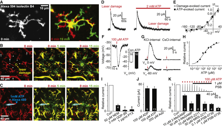
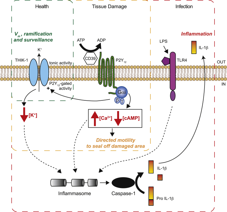
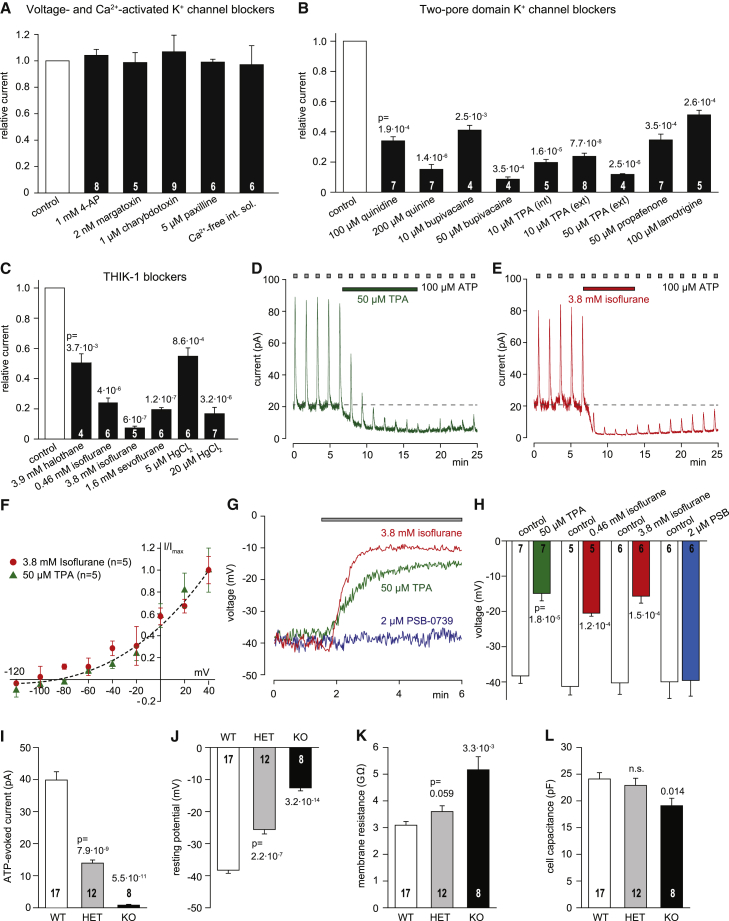
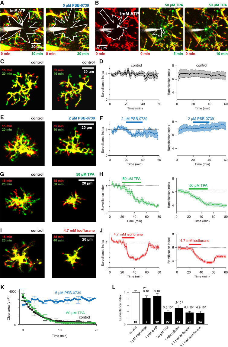
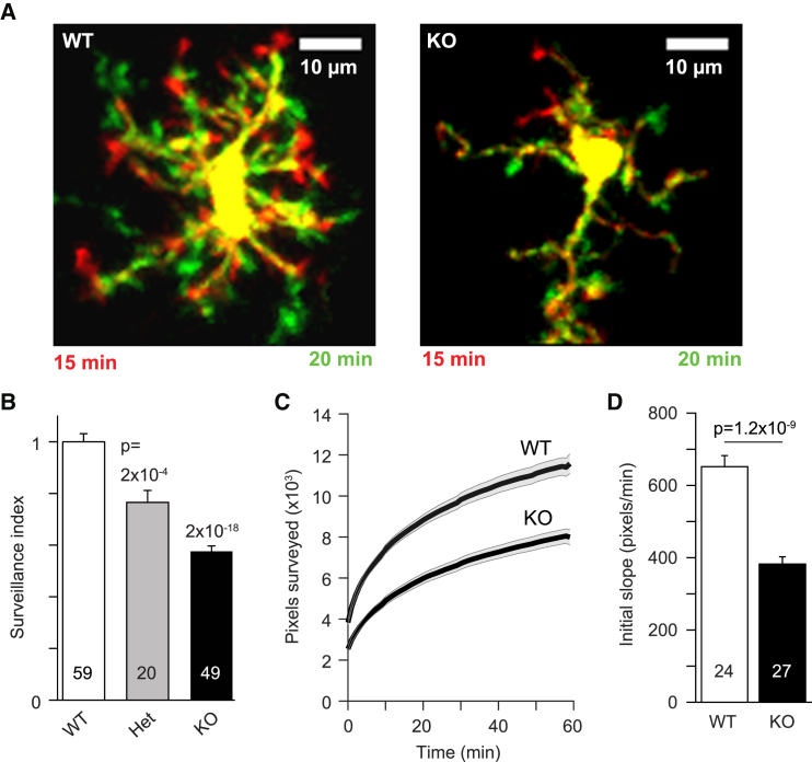
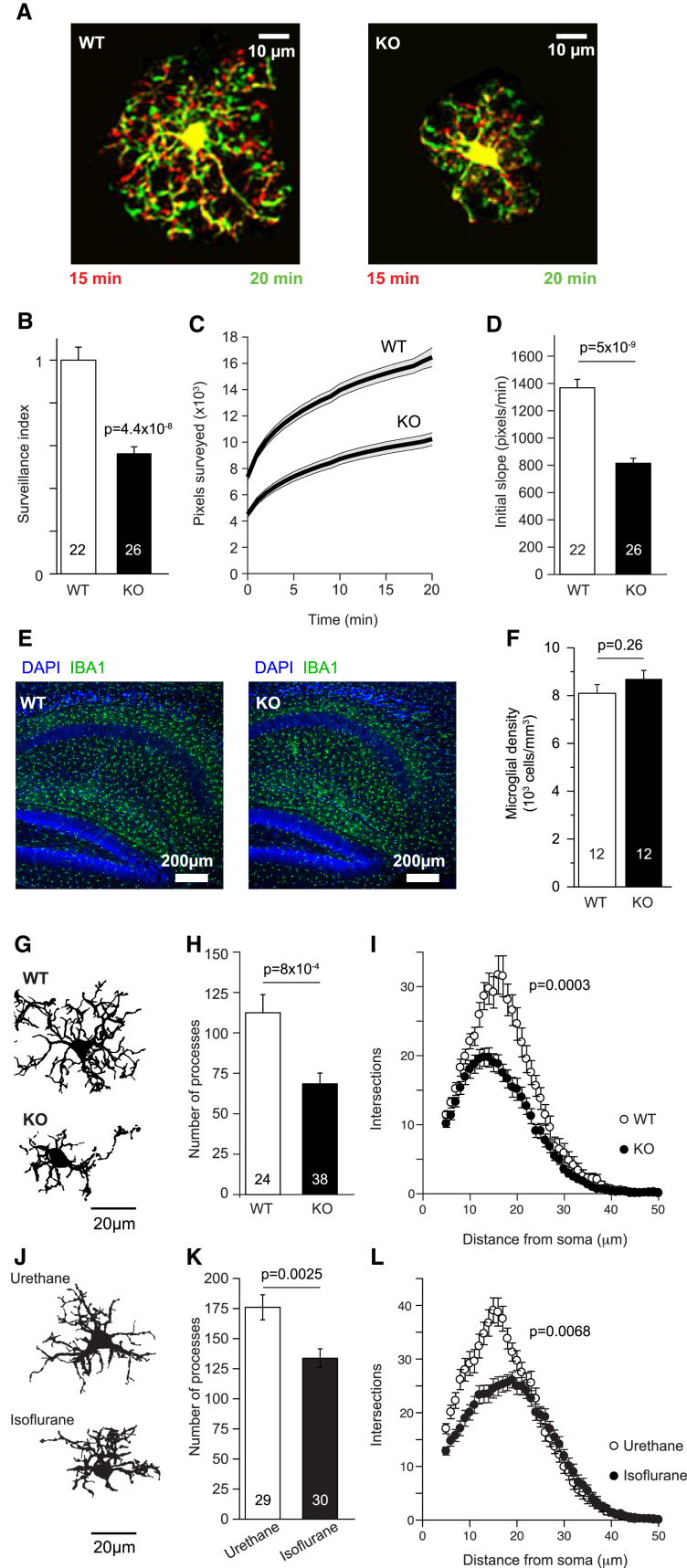
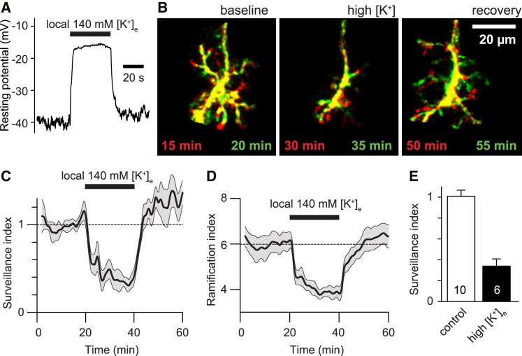
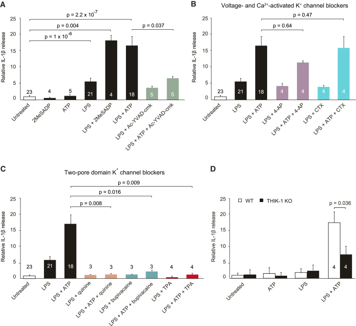
Microglia exhibit two modes of motility: they constantly extend and retract their processes to survey the brain, but they also send out targeted processes to envelop sites of tissue damage. We now show that these motility modes differ mechanistically. We identify the two-pore domain channel THIK-1 as the main K channel expressed in microglia in situ. THIK-1 is tonically active, and its activity is potentiated by P2Y receptors. Inhibiting THIK-1 function pharmacologically or by gene knockout depolarizes microglia, which decreases microglial ramification and thus reduces surveillance, whereas blocking P2Y receptors does not affect membrane potential, ramification, or surveillance. In contrast, process outgrowth to damaged tissue requires P2Y receptor activation but is unaffected by blocking THIK-1. Block of THIK-1 function also inhibits release of the pro-inflammatory cytokine interleukin-1β from activated microglia, consistent with K loss being needed for inflammasome assembly. Thus, microglial immune surveillance and cytokine release require THIK-1 channel activity.
它们不断地伸展和缩回其突起以监测大脑,但它们也会发出有针对性的突起来包围组织损伤部位。我们现在表明,这些运动模式在机制上有所不同。我们确定双孔域通道 THIK-1 是原位小胶质细胞中主要表达的 K 通道。THIK-1 持续活跃,其活性被 P2Y 受体增强。药理学抑制 THIK-1 功能或基因敲除会使小胶质细胞去极化,从而减少小胶质细胞的分支,从而减少监测,而阻断 P2Y 受体则不会影响膜电位、分支或监测。相比之下,向受损组织的突起生长需要 P2Y 受体的激活,但阻断 THIK-1 则不会影响突起生长。THIK-1 功能的阻断也抑制了激活的小胶质细胞中促炎细胞因子白细胞介素-1β的释放,这与炎症小体组装需要 K 丢失一致。因此,小胶质细胞的免疫监测和细胞因子释放需要 THIK-1 通道活性。