Henderson L E, Abdelmegeed M A, Yoo S H, Rhee S G, Zhu X, Smith M A, Nguyen R Q, Perry G, Song B J
Laboratory of Membrane Biochemistry and Biophysics, National Institute on Alcohol Abuse and Alcoholism, Bethesda, Maryland 20892-9410, USA.
Division of Life and Pharmaceutical Sciences, Ewha Womans University, Seoul, Korea.
Open Neurol J. 2017 Nov 16;11:48-58. doi: 10.2174/1874205X01711010048. eCollection 2017.
Despite increased neuronal death, senile plaques, and neurofibrillary tangles observed in patients suffering from Alzheimer's disease (AD), the detailed mechanism of cell death in AD is still poorly understood.
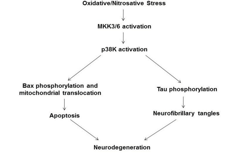
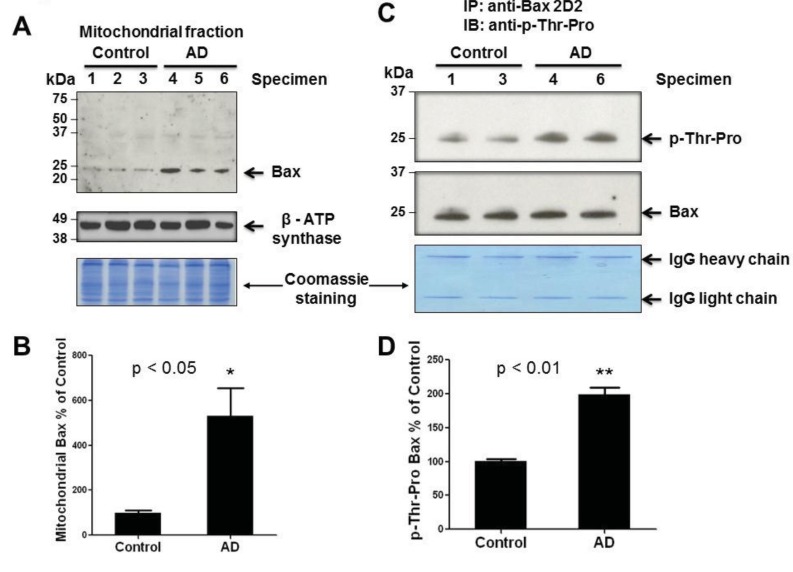
We hypothesized that p38 kinase activates and then phosphorylates Bax, leading to its translocation to mitochondria in AD brains compared to controls. The aim of this study was to investigate the role of p38 kinase in phosphorylation and sub-cellular localization of pro-apoptotic Bax in the frontal cortex of the brains from AD and control subjects. Increased oxidative stress in AD individuals compared to control was evaluated by measuring the levels of carbonylated proteins and oxidized peroxiredoxin, an antioxidant enzyme. The relative amounts of p38 kinase and phospho-Bax in mitochondria in AD brains and controls were determined by immunoblot analysis using the respective antibody against each protein following immunoprecipitation.
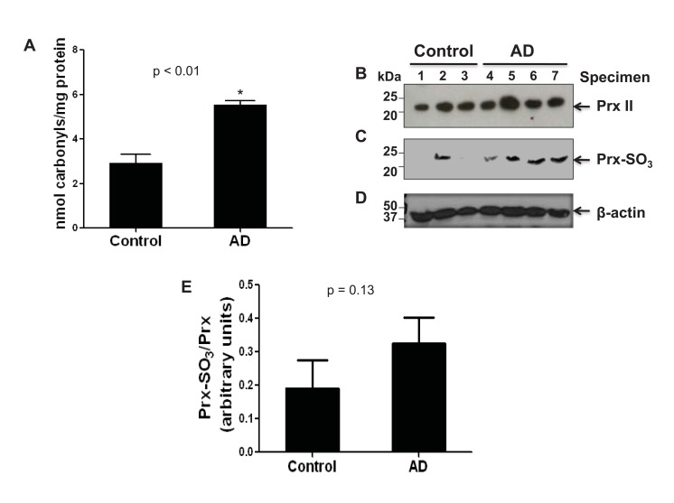
Our results showed that the levels of oxidized peroxiredoxin-SO and carbonylated proteins are significantly elevated in AD brains compared to controls, demonstrating the increased oxidative stress.
The amount of phospho-p38 kinase is increased in AD brains and the activated p38 kinase appears to phosphorylate Thr residue(s) of Bax, which leads to its mitochondrial translocation, contributing to apoptosis and ultimately, neurodegeneration.
尽管在阿尔茨海默病(AD)患者中观察到神经元死亡增加、老年斑和神经原纤维缠结,但AD中细胞死亡的详细机制仍知之甚少。
我们假设p38激酶被激活,然后使Bax磷酸化,导致其在AD脑中转位至线粒体,而对照组则不然。本研究的目的是调查p38激酶在AD和对照受试者大脑额叶皮质中促凋亡Bax的磷酸化和亚细胞定位中的作用。通过测量羰基化蛋白和氧化过氧化物酶(一种抗氧化酶)的水平,评估AD个体与对照组相比氧化应激的增加情况。在免疫沉淀后,使用针对每种蛋白质的相应抗体,通过免疫印迹分析确定AD脑和对照中线粒体中p38激酶和磷酸化Bax的相对含量。
我们的结果表明,与对照组相比,AD脑中氧化过氧化物酶-SO和羰基化蛋白的水平显著升高,表明氧化应激增加。
AD脑中磷酸化p38激酶的量增加,激活的p38激酶似乎使Bax的苏氨酸残基磷酸化,导致其线粒体转位,促成细胞凋亡并最终导致神经退行性变。