Menezes Rubens Alex de Oliveira, Gomes Margarete do Socorro Mendonça, Mendes Anapaula Martins, Couto Álvaro Augusto Ribeiro D' Almeida, Nacher Mathieu, Pimenta Tamirys Simão, Sousa Aline Collares Pinheiro de, Baptista Andrea Regina de Souza, Jesus Maria Izabel de, Enk Martin Johannes, Cunha Maristela Gomes, Machado Ricardo Luiz Dantas
Postgraduate Program in the Biology of Infectious and Parasitic Agents, Federal University of Pará (UFPA), Belém, Pará State, Brazil.
Laboratory of morphofunctional and parasitic studies with impact on health (LEMPIS), Federal University of Amapá (UNIFAP), Macapa, Amapá State, Brazil.
PLoS One. 2018 Jan 2;13(1):e0189958. doi: 10.1371/journal.pone.0189958. eCollection 2018.
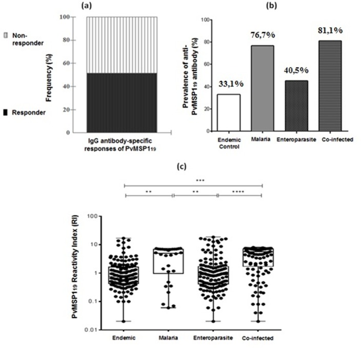
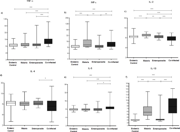
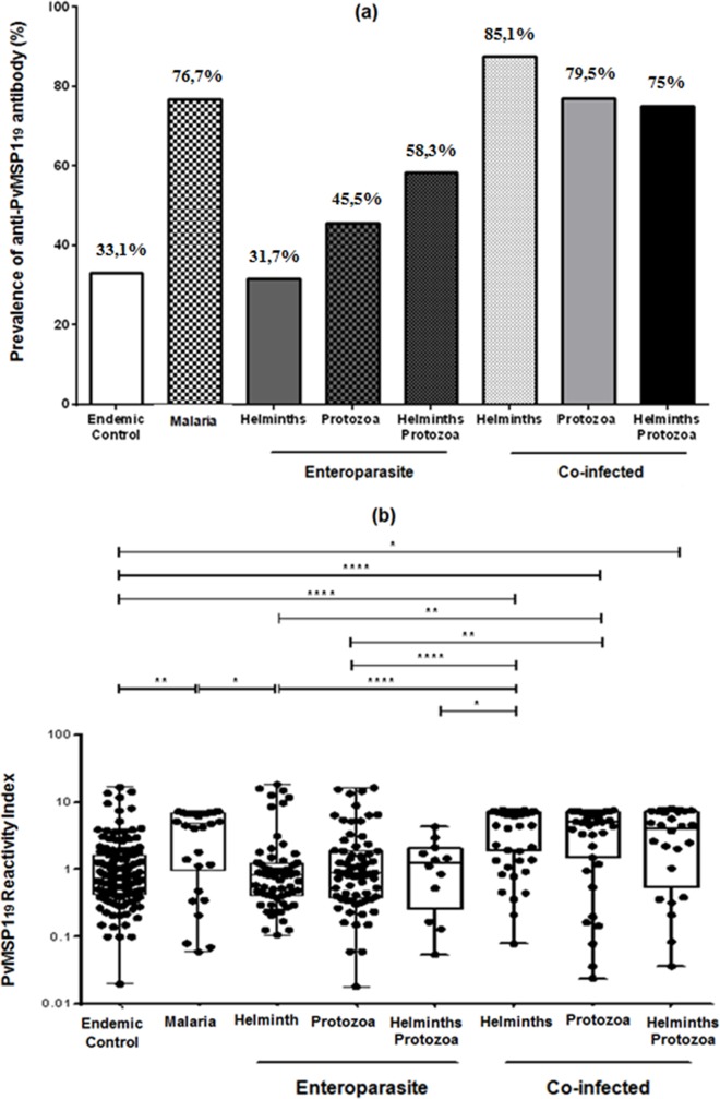
Malaria-enteroparasitic co-infections are known for their endemicity. Although they are prevalent, little is known about their epidemiology and effect on the immune response. This study evaluated the effect of enteroparasite co-infections with malaria caused by Plasmodium vivax in a border area between Brazil and French Guiana. The cross sectional study took place in Oiapoque, a municipality of Amapá, on the Amazon border. Malaria was diagnosed using thick blood smears, haemoglobin dosage by an automated method and coproparasitology by the Hoffman and Faust methods. The anti-PvMSP-119 IgG antibodies in the plasma were evaluated using ELISA and Th1 (IFN-γ, TNF-α and IL-2), and Th2 (IL-4, IL-5 and IL-10) cytokine counts were performed by flow cytometry. The participants were grouped into those that were monoinfected with vivax malaria (M), vivax malaria-enteroparasite co-infected (CI), monoinfected with enteroparasite (E) and endemic controls (EC), who were negative for both diseases. 441 individuals were included and grouped according to their infection status: [M 6.9% (30/441)], [Cl 26.5% (117/441)], [E 32.4% (143/441)] and [EC 34.2% (151/441)]. Males prevailed among the (M) 77% (23/30) and (CI) 60% (70/117) groups. There was a difference in haemoglobin levels among the different groups under study for [EC-E], [EC-Cl], [E-M] and [Cl-M], with (p < 0.01). Anaemia was expressed as a percentage between individuals [CI-EC (p < 0.05)]. In terms of parasitaemia, there were differences for the groups [CI-M (p < 0.05)]. Anti-PvMSP-119 antibodies were detected in 51.2% (226/441) of the population. The level of cytokines evaluation revealed a large variation in TNF-α and IL-10 concentrations in the co-infected group. In this study we did not observe any influence of coinfection on the acquisition of IgG antibodies against PvMSP119, as well as on the profile of the cytokines that characterize the Th1 and Th2 patterns. However, co-infection increased TNF-α and IL-10 levels.
疟疾与肠道寄生虫共感染以其地方性流行而闻名。尽管它们很普遍,但对其流行病学及其对免疫反应的影响却知之甚少。本研究评估了在巴西和法属圭亚那边境地区,间日疟原虫引起的疟疾与肠道寄生虫共感染的影响。这项横断面研究在亚马孙边境阿马帕州的奥亚波克市进行。使用厚血涂片诊断疟疾,通过自动方法测定血红蛋白含量,并采用霍夫曼法和福斯特法进行粪便寄生虫学检查。使用酶联免疫吸附测定法评估血浆中的抗间日疟原虫裂殖子表面蛋白119(PvMSP-119)IgG抗体,并通过流式细胞术检测Th1(干扰素-γ、肿瘤坏死因子-α和白细胞介素-2)和Th2(白细胞介素-4、白细胞介素-5和白细胞介素-10)细胞因子计数。参与者被分为单纯感染间日疟原虫(M)、间日疟原虫与肠道寄生虫共感染(CI)、单纯感染肠道寄生虫(E)和地方性对照(EC,两种疾病均为阴性)。根据感染状况纳入441人并分组:[M组6.9%(30/441)]、[CI组26.5%(117/441)]、[E组32.4%(143/441)]和[EC组34.2%(151/441)]。在(M)组的77%(23/30)和(CI)组的60%(70/117)中男性占多数。在[EC-E]、[EC-CI]、[E-M]和[CI-M]等不同研究组之间,血红蛋白水平存在差异(p<0.01)。贫血以个体间的百分比表示[CI-EC(p<0.05)]。就寄生虫血症而言,[CI-M]组存在差异(p<0.05)。在51.2%(226/441)的人群中检测到抗PvMSP-119抗体。细胞因子评估水平显示,共感染组中肿瘤坏死因子-α和白细胞介素-10浓度存在很大差异。在本研究中,我们未观察到共感染对获得抗PvMSP119 IgG抗体以及对表征Th1和Th2模式的细胞因子谱有任何影响。然而,共感染会增加肿瘤坏死因子-α和白细胞介素-10水平。