Lankford Karen L, Arroyo Edgardo J, Nazimek Katarzyna, Bryniarski Krzysztof, Askenase Philip W, Kocsis Jeffery D
Department of Neurology, Yale University School of Medicine, New Haven, Connecticut, United States of America.
Center for Neuroscience Regeneration Research, VA Connecticut Healthcare System, West Haven, Connecticut, United States of America.
PLoS One. 2018 Jan 2;13(1):e0190358. doi: 10.1371/journal.pone.0190358. eCollection 2018.
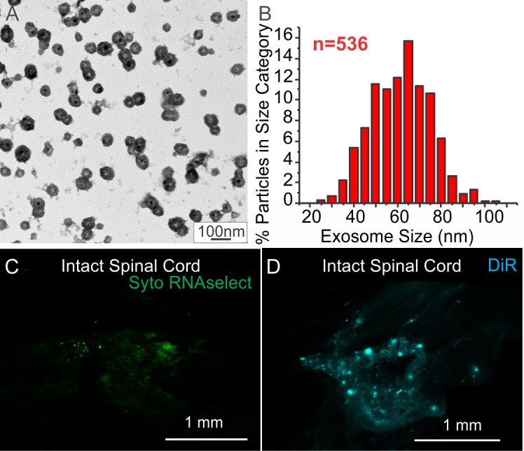
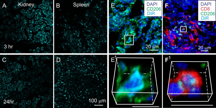
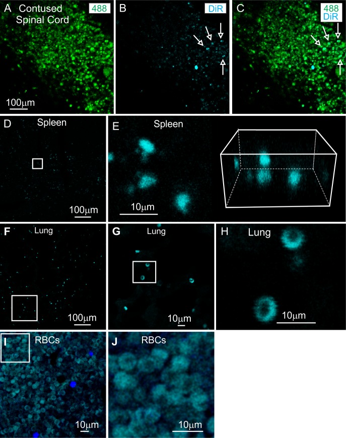
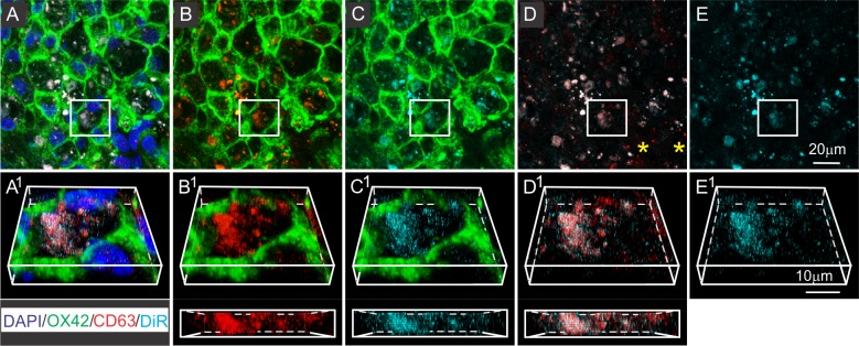
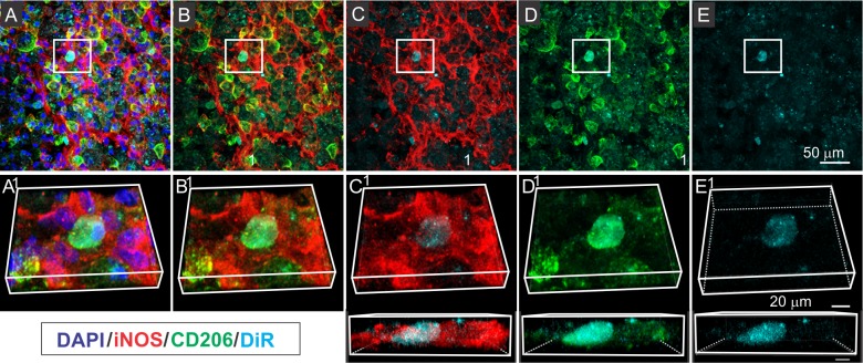
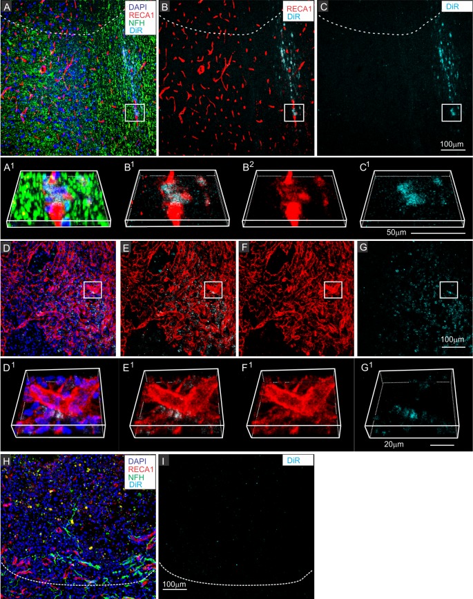
In a previous report we showed that intravenous infusion of bone marrow-derived mesenchymal stem cells (MSCs) improved functional recovery after contusive spinal cord injury (SCI) in the non-immunosuppressed rat, although the MSCs themselves were not detected at the spinal cord injury (SCI) site [1]. Rather, the MSCs lodged transiently in the lungs for about two days post-infusion. Preliminary studies and a recent report [2] suggest that the effects of intravenous (IV) infusion of MSCs could be mimicked by IV infusion of exosomes isolated from conditioned media of MSC cultures (MSCexos). In this study, we assessed the possible mechanism of MSCexos action on SCI by investigating the tissue distribution and cellular targeting of DiR fluorescent labeled MSCexos at 3 hours and 24 hours after IV infusion in rats with SCI. The IV delivered MSCexos were detected in contused regions of the spinal cord, but not in the noninjured region of the spinal cord, and were also detected in the spleen, which was notably reduced in weight in the SCI rat, compared to control animals. DiR "hotspots" were specifically associated with CD206-expressing M2 macrophages in the spinal cord and this was confirmed by co-localization with anti-CD63 antibodies labeling a tetraspanin characteristically expressed on exosomes. Our findings that MSCexos specifically target M2-type macrophages at the site of SCI, support the idea that extracellular vesicles, released by MSCs, may mediate at least some of the therapeutic effects of IV MSC administration.
在之前的一份报告中,我们表明,在非免疫抑制大鼠中,静脉输注骨髓间充质干细胞(MSCs)可改善挫伤性脊髓损伤(SCI)后的功能恢复,尽管在脊髓损伤(SCI)部位未检测到MSCs本身[1]。相反,MSCs在输注后约两天短暂驻留在肺部。初步研究和最近的一份报告[2]表明,静脉输注MSCs的效果可通过静脉输注从MSC培养条件培养基中分离的外泌体(MSCexos)来模拟。在本研究中,我们通过在SCI大鼠静脉输注后3小时和24小时研究DiR荧光标记的MSCexos的组织分布和细胞靶向,评估了MSCexos对SCI作用的可能机制。静脉输注的MSCexos在脊髓挫伤区域被检测到,但在脊髓未损伤区域未被检测到,并且在脾脏中也被检测到,与对照动物相比,SCI大鼠的脾脏重量明显减轻。DiR“热点”与脊髓中表达CD206的M2巨噬细胞特异性相关,这通过与标记外泌体上特征性表达的四跨膜蛋白的抗CD63抗体共定位得到证实。我们的研究结果表明,MSCexos在SCI部位特异性靶向M2型巨噬细胞,支持了MSCs释放的细胞外囊泡可能介导静脉输注MSC至少部分治疗效果的观点。