Marsman Gerben, von Richthofen Helen, Bulder Ingrid, Lupu Florea, Hazelzet Jan, Luken Brenda M, Zeerleder Sacha
Department of Immunopathology, Sanquin Research and Landsteiner Laboratory, Academic Medical Center, University of Amsterdam, Amsterdam, The Netherlands.
Cardiovascular Biology Research Program, Oklahoma Medical Research Foundation, Oklahoma City, OK.
Blood Adv. 2017 Nov 30;1(26):2491-2502. doi: 10.1182/bloodadvances.2017010959. eCollection 2017 Dec 12.
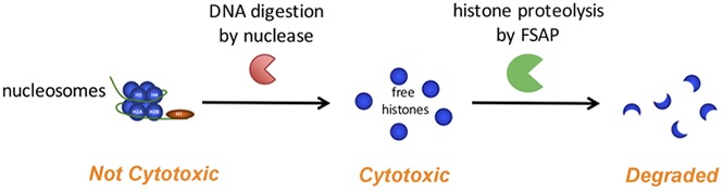
Circulating histones have been implicated as major mediators of inflammatory disease because of their strong cytotoxic effects. Histones form the protein core of nucleosomes; however, it is unclear whether histones and nucleosomes are equally cytotoxic. Several plasma proteins that neutralize histones are present in plasma. Importantly, factor VII-activating protease (FSAP) is activated upon contact with histones and subsequently proteolyzes histones. We aimed to determine the effect of FSAP on the cytotoxicity of both histones and nucleosomes. Indeed, FSAP protected against histone-induced cytotoxicity of cultured cells in vitro. Upon incubation of serum with histones, endogenous FSAP was activated and degraded histones, which also prevented cytotoxicity. Notably, histones as part of nucleosome complexes were not cytotoxic, whereas DNA digestion restored cytotoxicity. Histones in nucleosomes were inefficiently cleaved by FSAP, which resulted in limited cleavage of histone H3 and removal of the N-terminal tail. The specific isolation of either circulating nucleosomes or free histones from sera of challenged baboons or patients with meningococcal sepsis revealed that histone H3 was present in the form of nucleosomes, whereas free histone H3 was not detected. All samples showed signs of FSAP activation. Markedly, we observed that all histone H3 in nucleosomes from the patients with sepsis, and most histone H3 from the baboons, was N-terminally truncated, giving rise to a similarly sized protein fragment as through cleavage by FSAP. Taken together, our results suggest that DNA and FSAP jointly limit histone cytotoxicity and that free histone H3 does not circulate in appreciable concentrations in sepsis.
循环组蛋白因其强大的细胞毒性作用而被认为是炎症性疾病的主要介质。组蛋白构成核小体的蛋白质核心;然而,尚不清楚组蛋白和核小体的细胞毒性是否相同。血浆中存在几种中和组蛋白的血浆蛋白。重要的是,因子VII激活蛋白酶(FSAP)在与组蛋白接触时被激活,随后对组蛋白进行蛋白水解。我们旨在确定FSAP对组蛋白和核小体细胞毒性的影响。事实上,FSAP在体外可保护培养细胞免受组蛋白诱导的细胞毒性。血清与组蛋白孵育后,内源性FSAP被激活并降解组蛋白,这也可防止细胞毒性。值得注意的是,作为核小体复合物一部分的组蛋白没有细胞毒性,而DNA消化可恢复细胞毒性。核小体中的组蛋白被FSAP低效切割,导致组蛋白H3的切割有限且N端尾巴被去除。从受挑战的狒狒或脑膜炎球菌败血症患者的血清中特异性分离循环核小体或游离组蛋白,结果显示组蛋白H3以核小体形式存在,而未检测到游离组蛋白H3。所有样本均显示出FSAP激活的迹象。明显地,我们观察到败血症患者核小体中的所有组蛋白H3以及狒狒的大多数组蛋白H3在N端被截断,产生了与FSAP切割后大小相似的蛋白质片段。综上所述,我们的结果表明DNA和FSAP共同限制组蛋白的细胞毒性,并且在败血症中游离组蛋白H3不会以可观的浓度循环。