Teraphongphom Nutte, Kong Christina S, Warram Jason M, Rosenthal Eben L
Department of Otolaryngology-Head & Neck Surgery Stanford University, Stanford California U.S.A.
Department of Pathology Stanford University Stanford California U.S.A.
Laryngoscope Investig Otolaryngol. 2017 Oct 16;2(6):447-452. doi: 10.1002/lio2.84. eCollection 2017 Dec.
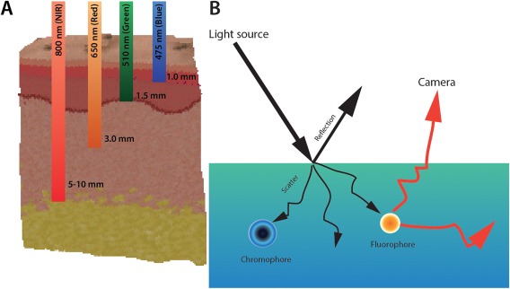
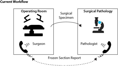
Although the agreed-upon standard is circumferential pathology analysis of the interface between the resected specimen and the patient, there is currently no consensus on the optimal methodology to achieve this in head and neck cancer specimens. This is most commonly conducted by either sampling the wound bed after resection or obtaining samples from the specimen. Regardless of the technique, only a fraction of the area of interest can be sampled due to the labor-intensive nature of frozen sections.
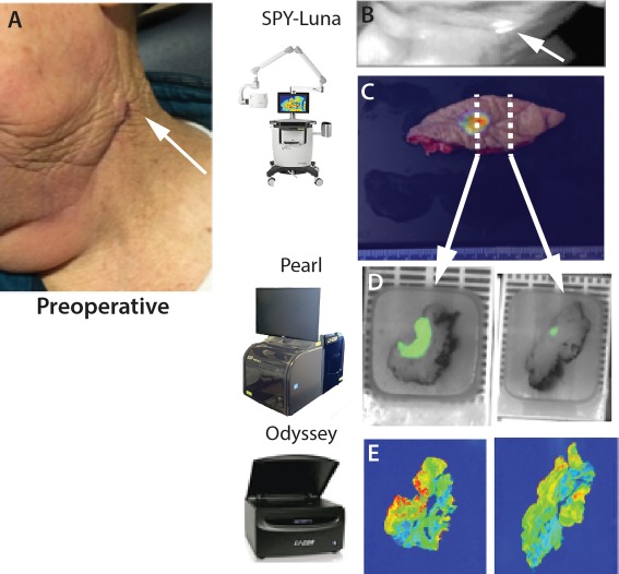
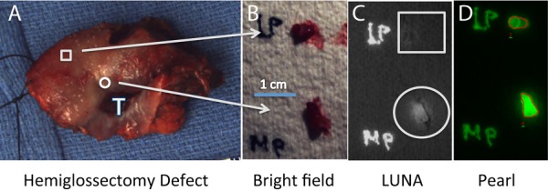
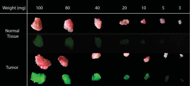
This review will cover and define the possible role for optical mapping of the surgical specimen using fluorescence imaging in head and neck cancer.
NA.
虽然公认的标准是对切除标本与患者之间的界面进行周向病理分析,但目前在头颈部癌标本中实现这一目标的最佳方法尚无共识。这最常见的做法是在切除后对伤口床进行采样或从标本中获取样本。无论采用何种技术,由于冰冻切片的劳动强度大,只能对感兴趣区域的一小部分进行采样。
本综述将涵盖并定义在头颈部癌中使用荧光成像对手术标本进行光学映射的可能作用。
无。