• 文献检索
  • 文档翻译
  • 深度研究
  • 学术资讯
  • Suppr Zotero 插件Zotero 插件
  • 邀请有礼
  • 套餐&价格
  • 历史记录
应用&插件
Suppr Zotero 插件Zotero 插件浏览器插件Mac 客户端Windows 客户端微信小程序
定价
高级版会员购买积分包购买API积分包
服务
文献检索文档翻译深度研究API 文档MCP 服务
关于我们
关于 Suppr公司介绍联系我们用户协议隐私条款
关注我们

Suppr 超能文献

核心技术专利:CN118964589B侵权必究
粤ICP备2023148730 号-1Suppr @ 2026

文献检索

告别复杂PubMed语法,用中文像聊天一样搜索,搜遍4000万医学文献。AI智能推荐,让科研检索更轻松。

立即免费搜索

文件翻译

保留排版,准确专业,支持PDF/Word/PPT等文件格式,支持 12+语言互译。

免费翻译文档

深度研究

AI帮你快速写综述,25分钟生成高质量综述,智能提取关键信息,辅助科研写作。

立即免费体验

术中分子成像用于评估口腔鳞状细胞癌的切缘情况

Intraoperative Molecular Imaging for Assessment of Peripheral Margins in Oral Squamous Cell Carcinoma.

作者信息

Fakurnejad Shayan, Krishnan Giri, van Keulen Stan, Nishio Naoki, Birkeland Andrew C, Baik Fred M, Kaplan Michael J, Colevas A Dimitrios, van den Berg Nynke S, Rosenthal Eben L, Martin Brock A

机构信息

Department of Otolaryngology - Head and Neck Surgery, Stanford University School of Medicine, Stanford, CA, United States.

The Department of Otorhinolaryngology, Head and Neck Surgery, The University of Adelaide, Woodville South, SA, Australia.

出版信息

Front Oncol. 2020 Jan 10;9:1476. doi: 10.3389/fonc.2019.01476. eCollection 2019.

DOI:10.3389/fonc.2019.01476
PMID:31998640
原文链接:https://pmc.ncbi.nlm.nih.gov/articles/PMC6965069/
Abstract

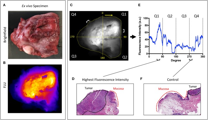
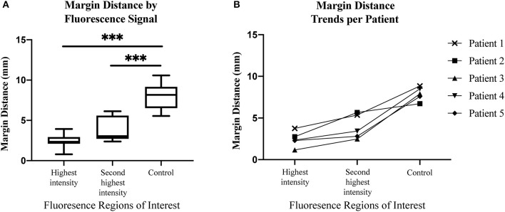
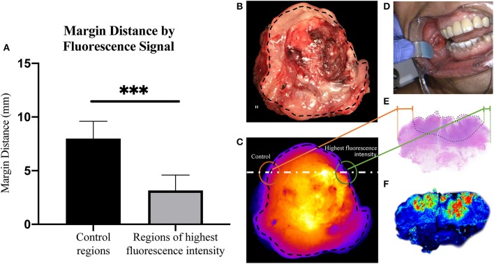
Complete surgical resection is the standard of care for treatment of oral cancer although the positive margin rate remains 15-30%. Tissue sampling from the resected specimen and from the wound bed for frozen section analysis (FSA) remains the mainstay for intraoperative margin assessment but is subject to sampling error and can require the processing of multiple samples. We sought to understand if an imaging strategy using a tumor-targeted fluorescently labeled antibody could accurately identify the closest peripheral margin on the mucosal surface of resected tumor specimen, so that this "sentinel margin" could be used to guide pathological sampling. Twenty-nine patients with oral squamous cell carcinoma scheduled for surgical resection were consented for the study and received systemic administration of a tumor-targeted fluorescently labeled antibody (Panitumumab IRDye800CW). After surgical resection, the tumor specimen was imaged using a closed-field fluorescent imaging device. Relevant pathological data was available for five patients on retrospective review. For each of these five patients, two regions of highest fluorescence intensity at the peripheral margin and one region of lowest fluorescence intensity were identified, and results were correlated with histology to determine if the region of highest fluorescence intensity along the mucosal margin (i.e., the sentinel margin) was truly the closest margin. Imaging acquisition of the mucosal surface of the specimen immediately after surgery took 30 s. In all of the specimens, the region of highest fluorescence at the specimen edge had a significantly smaller margin distance than other sampled regions. The average margin distance at the closest, "sentinel," margin was 3.2 mm compared to a margin distance of 8.0 mm at other regions ( < 0.0001). This proof-of-concept study suggests that, when combined with routine FSA, fluorescent specimen imaging can be used to identify the closest surgical margin on the specimen. This approach may reduce sampling error of intraoperative evaluation, which should ultimately improve the ability of the surgeon to identify the sentinel margin. This rapid sentinel margin identification improves the surgeon's orientation to areas most likely to be positive in the surgical wound bed and may expedite pathology workflow.

摘要

完整的手术切除是口腔癌治疗的标准治疗方法,尽管切缘阳性率仍为15%-30%。对切除标本和创面床进行组织取样以进行冰冻切片分析(FSA)仍然是术中切缘评估的主要方法,但存在取样误差,并且可能需要处理多个样本。我们试图了解使用肿瘤靶向荧光标记抗体的成像策略是否能够准确识别切除肿瘤标本黏膜表面最接近的外周切缘,以便这个“前哨切缘”能够用于指导病理取样。29例计划进行手术切除的口腔鳞状细胞癌患者同意参与本研究,并接受了肿瘤靶向荧光标记抗体(帕尼单抗IRDye800CW)的全身给药。手术切除后,使用封闭场荧光成像设备对肿瘤标本进行成像。回顾性分析时可获得5例患者的相关病理数据。对于这5例患者中的每一例,在周边切缘确定两个荧光强度最高的区域和一个荧光强度最低的区域,并将结果与组织学进行关联,以确定黏膜切缘处荧光强度最高的区域(即前哨切缘)是否真的是最接近的切缘。手术后立即对标本黏膜表面进行成像采集耗时30秒。在所有标本中,标本边缘荧光强度最高的区域与其他取样区域相比,切缘距离明显更小。最接近的“前哨”切缘的平均切缘距离为3.2毫米,而其他区域的切缘距离为8.0毫米(<0.0001)。这项概念验证研究表明,与常规FSA相结合时,荧光标本成像可用于识别标本上最接近的手术切缘。这种方法可能会减少术中评估的取样误差,最终应能提高外科医生识别前哨切缘的能力。这种快速的前哨切缘识别改善了外科医生对手术创面床中最可能呈阳性区域的定位,并可能加快病理工作流程。

https://cdn.ncbi.nlm.nih.gov/pmc/blobs/12f9/6965069/85aba556b2fb/fonc-09-01476-g0003.jpg
https://cdn.ncbi.nlm.nih.gov/pmc/blobs/12f9/6965069/2ee3fa46c98e/fonc-09-01476-g0001.jpg
https://cdn.ncbi.nlm.nih.gov/pmc/blobs/12f9/6965069/4da7f823209a/fonc-09-01476-g0002.jpg
https://cdn.ncbi.nlm.nih.gov/pmc/blobs/12f9/6965069/85aba556b2fb/fonc-09-01476-g0003.jpg
https://cdn.ncbi.nlm.nih.gov/pmc/blobs/12f9/6965069/2ee3fa46c98e/fonc-09-01476-g0001.jpg
https://cdn.ncbi.nlm.nih.gov/pmc/blobs/12f9/6965069/4da7f823209a/fonc-09-01476-g0002.jpg
https://cdn.ncbi.nlm.nih.gov/pmc/blobs/12f9/6965069/85aba556b2fb/fonc-09-01476-g0003.jpg

相似文献

1
Intraoperative Molecular Imaging for Assessment of Peripheral Margins in Oral Squamous Cell Carcinoma.术中分子成像用于评估口腔鳞状细胞癌的切缘情况
Front Oncol. 2020 Jan 10;9:1476. doi: 10.3389/fonc.2019.01476. eCollection 2019.
2
Fluorescent Molecular Imaging Can Improve Intraoperative Sentinel Margin Detection in Oral Squamous Cell Carcinoma.荧光分子成像可提高口腔鳞状细胞癌术中前哨切缘检测的准确性。
J Nucl Med. 2022 Aug;63(8):1162-1168. doi: 10.2967/jnumed.121.262235. Epub 2022 Jan 13.
3
The Sentinel Margin: Intraoperative Specimen Mapping Using Relative Fluorescence Intensity.前哨切缘:利用相对荧光强度进行术中标本定位
Clin Cancer Res. 2019 Aug 1;25(15):4656-4662. doi: 10.1158/1078-0432.CCR-19-0319. Epub 2019 May 29.
4
Frozen Section Analysis and Real-Time Magnetic Resonance Imaging of Surgical Specimen Oriented on 3D Printed Tongue Model to Assess Surgical Margins in Oral Tongue Carcinoma: Preliminary Results.基于3D打印舌模型的手术标本冷冻切片分析及实时磁共振成像评估口腔舌癌手术切缘:初步结果
Front Oncol. 2021 Dec 9;11:735002. doi: 10.3389/fonc.2021.735002. eCollection 2021.
5
Rapid, non-invasive fluorescence margin assessment: Optical specimen mapping in oral squamous cell carcinoma.快速、无创的荧光边缘评估:口腔鳞状细胞癌的光学标本绘图。
Oral Oncol. 2019 Jan;88:58-65. doi: 10.1016/j.oraloncology.2018.11.012. Epub 2018 Nov 21.
6
Determination of Tumor Margins with Surgical Specimen Mapping Using Near-Infrared Fluorescence.使用近红外荧光对手术标本进行肿瘤边界定位。
Cancer Res. 2018 Sep 1;78(17):5144-5154. doi: 10.1158/0008-5472.CAN-18-0878. Epub 2018 Jul 2.
7
Early Oral Tongue Squamous Cell Carcinoma: Sampling of Margins From Tumor Bed and Worse Local Control.早期口腔舌鳞状细胞癌:肿瘤床边缘的取样与较差的局部控制。
JAMA Otolaryngol Head Neck Surg. 2015 Dec;141(12):1104-10. doi: 10.1001/jamaoto.2015.1351.
8
Independent Predictors of Prognosis Based on Oral Cavity Squamous Cell Carcinoma Surgical Margins.基于口腔鳞状细胞癌手术切缘的预后独立预测因子。
Otolaryngol Head Neck Surg. 2018 Oct;159(4):675-682. doi: 10.1177/0194599818773070. Epub 2018 May 8.
9
Intraoperative Margin Assessment in Head and Neck Cancer: A Case of Misuse and Abuse?头颈部癌症的术中切缘评估:误用和滥用的案例?
Head Neck Pathol. 2020 Jun;14(2):291-302. doi: 10.1007/s12105-019-01121-2. Epub 2020 Mar 2.
10
Beneath the surface: A systematic review on intraoperative imaging techniques for deep margin assessment in oral squamous cell carcinoma.表面之下:口腔鳞状细胞癌术中评估深层切缘的影像学技术系统评价。
Oral Oncol. 2024 Jun;153:106823. doi: 10.1016/j.oraloncology.2024.106823. Epub 2024 May 2.

引用本文的文献

1
Fluorescence Imaging-Assessed Surgical Margin Detection in Head and Neck Oncology by Passive and Active Targeting.通过被动和主动靶向的荧光成像评估头颈部肿瘤手术切缘
Mol Diagn Ther. 2025 May 8. doi: 10.1007/s40291-025-00781-x.
2
Tumour Marker Expression in Head and Neck Malignancies to Identify Potential Targets for Intraoperative Molecular Near-Infrared Imaging.头颈部恶性肿瘤中肿瘤标志物的表达,以确定术中分子近红外成像的潜在靶点。
Mol Diagn Ther. 2024 Nov;28(6):811-820. doi: 10.1007/s40291-024-00742-w. Epub 2024 Sep 9.
3
Interim Phase II Results Using Panitumumab-IRDye800CW during Transoral Robotic Surgery in Patients with Oropharyngeal Cancer.

本文引用的文献

1
The Sentinel Margin: Intraoperative Specimen Mapping Using Relative Fluorescence Intensity.前哨切缘:利用相对荧光强度进行术中标本定位
Clin Cancer Res. 2019 Aug 1;25(15):4656-4662. doi: 10.1158/1078-0432.CCR-19-0319. Epub 2019 May 29.
2
Determination of Tumor Margins with Surgical Specimen Mapping Using Near-Infrared Fluorescence.使用近红外荧光对手术标本进行肿瘤边界定位。
Cancer Res. 2018 Sep 1;78(17):5144-5154. doi: 10.1158/0008-5472.CAN-18-0878. Epub 2018 Jul 2.
3
Safety of panitumumab-IRDye800CW and cetuximab-IRDye800CW for fluorescence-guided surgical navigation in head and neck cancers.
口咽癌患者经口机器人手术中使用 Panitumumab-IRDye800CW 的中期 II 期结果。
Clin Cancer Res. 2024 Sep 13;30(18):4016-4028. doi: 10.1158/1078-0432.CCR-24-0940.
4
Infrared Fluorescence-guided Surgery for Tumor and Metastatic Lymph Node Detection in Head and Neck Cancer.头颈部癌症中肿瘤和转移性淋巴结的红外荧光引导手术。
Radiol Imaging Cancer. 2024 Jul;6(4):e230178. doi: 10.1148/rycan.230178.
5
Surgical margins in head and neck squamous cell carcinoma: A narrative review.头颈部鳞状细胞癌的手术切缘:一篇叙述性综述。
Int J Surg. 2024 Jun 1;110(6):3680-3700. doi: 10.1097/JS9.0000000000001306.
6
Fluorescence molecular optomic signatures improve identification of tumors in head and neck specimens.荧光分子光学特征改善头颈部标本中肿瘤的识别。
Front Med Technol. 2023 Feb 15;5:1009638. doi: 10.3389/fmedt.2023.1009638. eCollection 2023.
7
Intraoperative Molecular Imaging of Lung Cancer: A Review.肺癌术中分子影像学:综述
Surg Oncol Clin N Am. 2022 Oct;31(4):685-693. doi: 10.1016/j.soc.2022.06.006. Epub 2022 Sep 27.
8
Validation of a surgical training model containing indocyanine green for near-infrared fluorescence imaging.包含吲哚菁绿用于近红外荧光成像的手术训练模型的验证
Laryngoscope Investig Otolaryngol. 2022 Jul 8;7(4):1011-1017. doi: 10.1002/lio2.858. eCollection 2022 Aug.
9
Preclinical Evaluation of an Activity-Based Probe for Intraoperative Imaging of Esophageal Cancer.基于活性的探针用于食管癌术中成像的临床前评估。
Mol Imaging. 2022 Jul 14;2022:5447290. doi: 10.1155/2022/5447290. eCollection 2022.
10
Three-Dimensional Near-Infrared Specimen Mapping Can Identify the Distance from the Tumor to the Surgical Margin During Resection of Pulmonary Ground Glass Opacities.三维近红外标本测绘可在切除肺部磨玻璃密度影时确定肿瘤与手术切缘的距离。
Mol Imaging Biol. 2023 Feb;25(1):203-211. doi: 10.1007/s11307-022-01750-0. Epub 2022 Jul 13.
头颈部癌症荧光引导手术导航中 panitumumab-IRDye800CW 和 cetuximab-IRDye800CW 的安全性。
Theranostics. 2018 Mar 28;8(9):2488-2495. doi: 10.7150/thno.24487. eCollection 2018.
4
Positive Surgical Margins in the 10 Most Common Solid Cancers.10 种最常见实体瘤中的阳性手术切缘。
Sci Rep. 2018 Apr 9;8(1):5686. doi: 10.1038/s41598-018-23403-5.
5
Fluorescence-guided surgery of cancer: applications, tools and perspectives.荧光引导手术治疗癌症:应用、工具和前景。
Curr Opin Chem Biol. 2018 Aug;45:64-72. doi: 10.1016/j.cbpa.2018.03.002. Epub 2018 Mar 23.
6
In-vivo optical imaging in head and neck oncology: basic principles, clinical applications and future directions.头颈部肿瘤的体内光学成像:基本原理、临床应用及未来方向。
Int J Oral Sci. 2018 Mar 18;10(2):10. doi: 10.1038/s41368-018-0011-4.
7
Optical molecular imaging for tumor detection and image-guided surgery.肿瘤检测和图像引导手术中的光学分子成像。
Biomaterials. 2018 Mar;157:62-75. doi: 10.1016/j.biomaterials.2017.12.002. Epub 2017 Dec 5.
8
Deep convolutional neural networks for classifying head and neck cancer using hyperspectral imaging.使用高光谱成像技术进行头颈部癌症分类的深度卷积神经网络。
J Biomed Opt. 2017 Jun 1;22(6):60503. doi: 10.1117/1.JBO.22.6.060503.
9
A Proposal to Redefine Close Surgical Margins in Squamous Cell Carcinoma of the Oral Tongue.关于重新定义舌鳞状细胞癌手术切缘的提议。
JAMA Otolaryngol Head Neck Surg. 2017 Jun 1;143(6):555-560. doi: 10.1001/jamaoto.2016.4238.
10
Oncologic Procedures Amenable to Fluorescence-guided Surgery.适用于荧光引导手术的肿瘤学手术程序。
Ann Surg. 2017 Jul;266(1):36-47. doi: 10.1097/SLA.0000000000002127.