• 文献检索
  • 文档翻译
  • 深度研究
  • 学术资讯
  • Suppr Zotero 插件Zotero 插件
  • 邀请有礼
  • 套餐&价格
  • 历史记录
应用&插件
Suppr Zotero 插件Zotero 插件浏览器插件Mac 客户端Windows 客户端微信小程序
定价
高级版会员购买积分包购买API积分包
服务
文献检索文档翻译深度研究API 文档MCP 服务
关于我们
关于 Suppr公司介绍联系我们用户协议隐私条款
关注我们

Suppr 超能文献

核心技术专利:CN118964589B侵权必究
粤ICP备2023148730 号-1Suppr @ 2026

文献检索

告别复杂PubMed语法,用中文像聊天一样搜索,搜遍4000万医学文献。AI智能推荐,让科研检索更轻松。

立即免费搜索

文件翻译

保留排版,准确专业,支持PDF/Word/PPT等文件格式,支持 12+语言互译。

免费翻译文档

深度研究

AI帮你快速写综述,25分钟生成高质量综述,智能提取关键信息,辅助科研写作。

立即免费体验

中重度创伤性脑损伤后脑容量进行性损失的空间模式。

Spatial patterns of progressive brain volume loss after moderate-severe traumatic brain injury.

机构信息

Computational, Cognitive and Clinical Neuroimaging Laboratory, Imperial College London, Division of Brain Sciences, Hammersmith Hospital, London, UK.

出版信息

Brain. 2018 Mar 1;141(3):822-836. doi: 10.1093/brain/awx354.

DOI:10.1093/brain/awx354
PMID:29309542
原文链接:https://pmc.ncbi.nlm.nih.gov/articles/PMC5837530/
Abstract

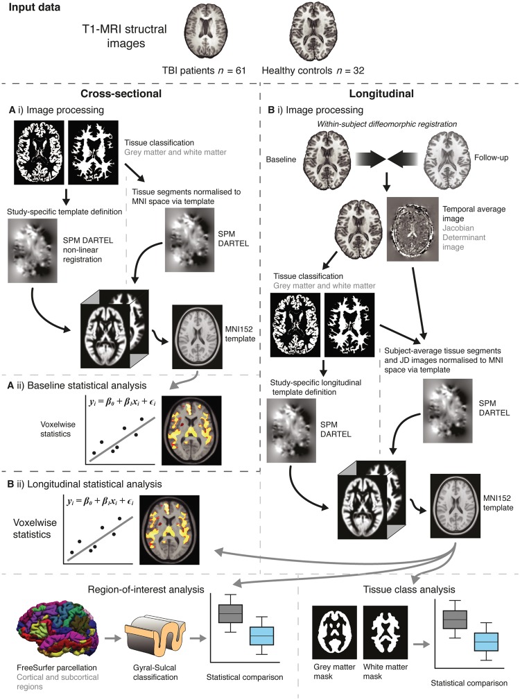
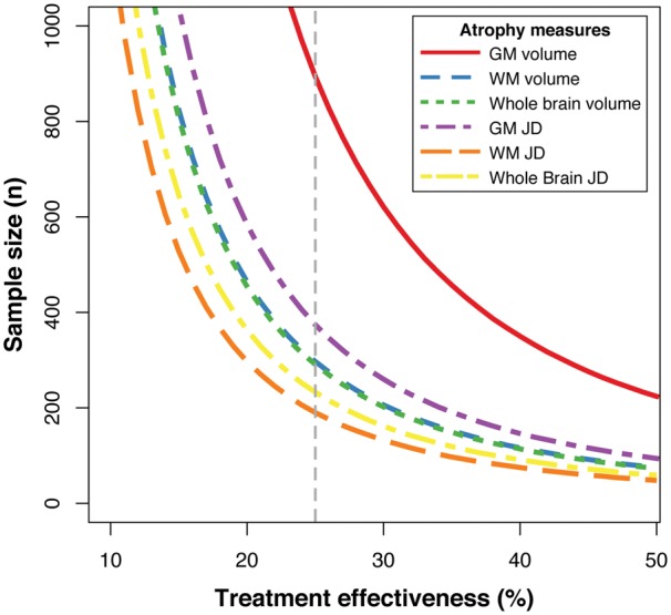
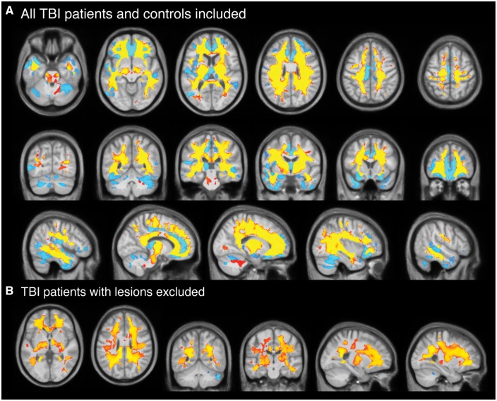
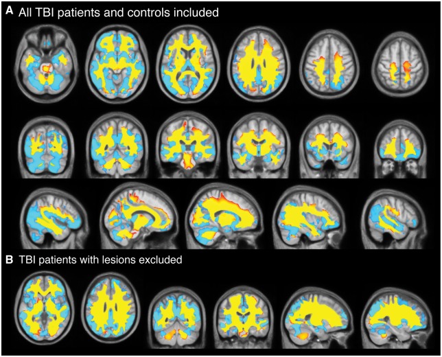
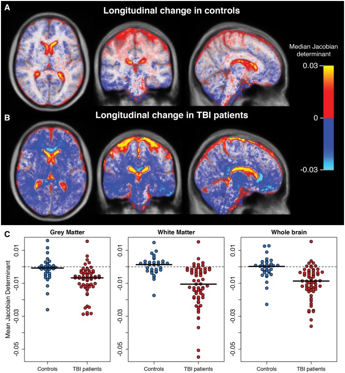
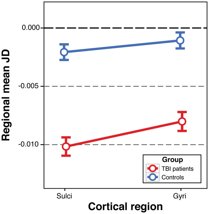
Traumatic brain injury leads to significant loss of brain volume, which continues into the chronic stage. This can be sensitively measured using volumetric analysis of MRI. Here we: (i) investigated longitudinal patterns of brain atrophy; (ii) tested whether atrophy is greatest in sulcal cortical regions; and (iii) showed how atrophy could be used to power intervention trials aimed at slowing neurodegeneration. In 61 patients with moderate-severe traumatic brain injury (mean age = 41.55 years ± 12.77) and 32 healthy controls (mean age = 34.22 years ± 10.29), cross-sectional and longitudinal (1-year follow-up) brain structure was assessed using voxel-based morphometry on T1-weighted scans. Longitudinal brain volume changes were characterized using a novel neuroimaging analysis pipeline that generates a Jacobian determinant metric, reflecting spatial warping between baseline and follow-up scans. Jacobian determinant values were summarized regionally and compared with clinical and neuropsychological measures. Patients with traumatic brain injury showed lower grey and white matter volume in multiple brain regions compared to controls at baseline. Atrophy over 1 year was pronounced following traumatic brain injury. Patients with traumatic brain injury lost a mean (± standard deviation) of 1.55% ± 2.19 of grey matter volume per year, 1.49% ± 2.20 of white matter volume or 1.51% ± 1.60 of whole brain volume. Healthy controls lost 0.55% ± 1.13 of grey matter volume and gained 0.26% ± 1.11 of white matter volume; equating to a 0.22% ± 0.83 reduction in whole brain volume. Atrophy was greatest in white matter, where the majority (84%) of regions were affected. This effect was independent of and substantially greater than that of ageing. Increased atrophy was also seen in cortical sulci compared to gyri. There was no relationship between atrophy and time since injury or age at baseline. Atrophy rates were related to memory performance at the end of the follow-up period, as well as to changes in memory performance, prior to multiple comparison correction. In conclusion, traumatic brain injury results in progressive loss of brain tissue volume, which continues for many years post-injury. Atrophy is most prominent in the white matter, but is also more pronounced in cortical sulci compared to gyri. These findings suggest the Jacobian determinant provides a method of quantifying brain atrophy following a traumatic brain injury and is informative in determining the long-term neurodegenerative effects after injury. Power calculations indicate that Jacobian determinant images are an efficient surrogate marker in clinical trials of neuroprotective therapeutics.

摘要

创伤性脑损伤导致大脑体积显著丧失,这种情况会持续到慢性阶段。这可以通过 MRI 的容积分析进行敏感测量。在这里,我们:(i) 研究了脑萎缩的纵向模式;(ii) 测试了萎缩是否在脑沟皮质区域最为严重;(iii) 展示了萎缩如何用于为旨在减缓神经退行性变的干预试验提供动力。在 61 名中度至重度创伤性脑损伤患者(平均年龄=41.55 岁±12.77 岁)和 32 名健康对照者(平均年龄=34.22 岁±10.29 岁)中,使用基于体素的形态测量学对 T1 加权扫描进行了横断面和纵向(1 年随访)脑结构评估。使用一种新的神经影像学分析管道对纵向脑体积变化进行了特征描述,该管道生成了雅可比行列式度量,反映了基线和随访扫描之间的空间变形。雅可比行列式值在区域上进行了总结,并与临床和神经心理学测量进行了比较。与基线相比,创伤性脑损伤患者在多个脑区的灰质和白质体积均较低。创伤性脑损伤后 1 年的萎缩明显。创伤性脑损伤患者每年平均损失 1.55%±2.19%的灰质体积、1.49%±2.20%的白质体积或 1.51%±1.60%的全脑体积。健康对照者每年损失 0.55%±1.13%的灰质体积,增加 0.26%±1.11%的白质体积;相当于全脑体积减少 0.22%±0.83%。萎缩最严重的是白质,其中 84%的区域受到影响。这种影响独立于且大大超过了年龄的影响。与脑回相比,皮质脑沟也出现了更多的萎缩。萎缩与损伤后时间或基线时的年龄无关。萎缩率与随访结束时的记忆表现有关,也与记忆表现的变化有关,这是在进行多次比较校正之前。总之,创伤性脑损伤会导致脑组织体积的进行性丧失,这种情况会持续多年。萎缩最明显的是白质,但与脑回相比,皮质脑沟更为明显。这些发现表明,雅可比行列式提供了一种量化创伤性脑损伤后脑萎缩的方法,对于确定损伤后的长期神经退行性效应具有信息性。功效计算表明,雅可比行列式图像是神经保护治疗临床试验中有效的替代标志物。

https://cdn.ncbi.nlm.nih.gov/pmc/blobs/2336/5837530/479382fc6dad/awx354f6.jpg
https://cdn.ncbi.nlm.nih.gov/pmc/blobs/2336/5837530/8bb8243cfb58/awx354f1.jpg
https://cdn.ncbi.nlm.nih.gov/pmc/blobs/2336/5837530/7515977439af/awx354f2.jpg
https://cdn.ncbi.nlm.nih.gov/pmc/blobs/2336/5837530/3168fbc2f170/awx354f3.jpg
https://cdn.ncbi.nlm.nih.gov/pmc/blobs/2336/5837530/9c2c6ca63f6b/awx354f4.jpg
https://cdn.ncbi.nlm.nih.gov/pmc/blobs/2336/5837530/4abb5a88efae/awx354f5.jpg
https://cdn.ncbi.nlm.nih.gov/pmc/blobs/2336/5837530/479382fc6dad/awx354f6.jpg

相似文献

1
Spatial patterns of progressive brain volume loss after moderate-severe traumatic brain injury.中重度创伤性脑损伤后脑容量进行性损失的空间模式。
Brain. 2018 Mar 1;141(3):822-836. doi: 10.1093/brain/awx354.
2
Diffuse axonal injury predicts neurodegeneration after moderate-severe traumatic brain injury.弥漫性轴索损伤预测中重度创伤性脑损伤后的神经退行性变。
Brain. 2020 Dec 1;143(12):3685-3698. doi: 10.1093/brain/awaa316.
3
Spatiotemporal profile of atrophy in the first year following moderate-severe traumatic brain injury.中重度创伤性脑损伤后第一年的萎缩时空分布特征。
Hum Brain Mapp. 2023 Sep;44(13):4692-4709. doi: 10.1002/hbm.26410. Epub 2023 Jul 3.
4
Serum NfL and GFAP as biomarkers of progressive neurodegeneration in TBI.血清 NfL 和 GFAP 作为 TBI 进行性神经退行性变的生物标志物。
Alzheimers Dement. 2024 Jul;20(7):4663-4676. doi: 10.1002/alz.13898. Epub 2024 May 28.
5
Brain Neurodegeneration in the Chronic Stage of the Survivors from Severe Non-Missile Traumatic Brain Injury: A Voxel-Based Morphometry Within-Group at One versus Nine Years from a Head Injury.严重非弹式颅脑创伤幸存者慢性期的脑神经退行性变:颅脑损伤后 1 年与 9 年的基于体素的形态计量学比较。
J Neurotrauma. 2021 Jan 15;38(2):283-290. doi: 10.1089/neu.2020.7203. Epub 2020 Nov 3.
6
Neuro-cognitive mechanisms of simultanagnosia in patients with posterior cortical atrophy.后部皮质萎缩患者同时失认症的神经认知机制。
Brain. 2016 Dec;139(Pt 12):3267-3280. doi: 10.1093/brain/aww235. Epub 2016 Oct 3.
7
Brain volume abnormalities and clinical outcomes following paediatric traumatic brain injury.儿童创伤性脑损伤后脑容量异常与临床结局。
Brain. 2022 Aug 27;145(8):2920-2934. doi: 10.1093/brain/awac130.
8
Post-acute blood biomarkers and disease progression in traumatic brain injury.创伤性脑损伤的急性后期血液生物标志物与疾病进展。
Brain. 2022 Jun 30;145(6):2064-2076. doi: 10.1093/brain/awac126.
9
Neuroanatomical correlates of prion disease progression - a 3T longitudinal voxel-based morphometry study.朊病毒病进展的神经解剖学关联——一项基于3T纵向体素的形态测量学研究。
Neuroimage Clin. 2016 Nov 2;13:89-96. doi: 10.1016/j.nicl.2016.10.021. eCollection 2017.
10
Predicting cognitive decline in multiple sclerosis: a 5-year follow-up study.多发性硬化症认知能力下降的预测:一项 5 年随访研究。
Brain. 2018 Sep 1;141(9):2605-2618. doi: 10.1093/brain/awy202.

引用本文的文献

1
Longitudinal Brain Structural, Neurochemical, and Behavioral Changes Following Traumatic Brain Injury in Immature Rat Brain with ALCAR Treatment.用ALCAR治疗的未成熟大鼠脑创伤性脑损伤后的纵向脑结构、神经化学和行为变化
Neurochem Res. 2025 Jul 30;50(4):256. doi: 10.1007/s11064-025-04494-9.
2
Distinct clinical phenotypes and their neuroanatomic correlates in chronic traumatic brain injury.慢性创伤性脑损伤的不同临床表型及其神经解剖学关联
Brain Commun. 2025 Jun 6;7(3):fcaf216. doi: 10.1093/braincomms/fcaf216. eCollection 2025.
3
Neuroimaging Correlates of Traumatic Brain Injury in an Older Community-Dwelling Population: The Atherosclerosis Risk in Communities Study.

本文引用的文献

1
Comparison of multiple tau-PET measures as biomarkers in aging and Alzheimer's disease.比较多种 tau-PET 指标作为衰老和阿尔茨海默病的生物标志物。
Neuroimage. 2017 Aug 15;157:448-463. doi: 10.1016/j.neuroimage.2017.05.058. Epub 2017 Jun 3.
2
The association between tau PET and retrospective cortical thinning in clinically normal elderly.tau PET 与临床正常老年人皮质变薄的相关性。
Neuroimage. 2017 Aug 15;157:612-622. doi: 10.1016/j.neuroimage.2017.05.049. Epub 2017 May 22.
3
Computational modelling of traumatic brain injury predicts the location of chronic traumatic encephalopathy pathology.
老年社区居住人群创伤性脑损伤的神经影像学关联:社区动脉粥样硬化风险研究
Neurology. 2025 Apr 22;104(8):e213506. doi: 10.1212/WNL.0000000000213506. Epub 2025 Apr 4.
4
Distinct clinical phenotypes and their neuroanatomic correlates in chronic traumatic brain injury.慢性创伤性脑损伤的不同临床表型及其神经解剖学关联
medRxiv. 2025 Jan 29:2025.01.27.25321200. doi: 10.1101/2025.01.27.25321200.
5
Changes in Hippocampal Volume after Traumatic Brain Injury (TBI).创伤性脑损伤(TBI)后海马体积的变化。
Res Sq. 2024 Dec 4:rs.3.rs-5390622. doi: 10.21203/rs.3.rs-5390622/v1.
6
Correction: Microglia are essential for tissue contraction in wound closure after brain injury in zebrafish larvae.更正:在斑马鱼幼体脑损伤后的伤口闭合过程中,小胶质细胞对于组织收缩至关重要。
Life Sci Alliance. 2024 Nov 25;8(2). doi: 10.26508/lsa.202403129. Print 2025 Feb.
7
Viral-mediated increased hippocampal neurogranin modulate synapses at one month in a rat model of controlled cortical impact.病毒介导的海马神经颗粒蛋白增加调节控制皮质撞击大鼠模型中一个月时的突触。
Sci Rep. 2024 Nov 22;14(1):28998. doi: 10.1038/s41598-024-77682-2.
8
The Effects of Early Life History of TBI on the Progression of Normal Brain Aging with Implications for Increased Dementia Risk.脑创伤后早期生活史对正常大脑老化进展的影响及其对痴呆风险增加的影响。
Adv Neurobiol. 2024;42:119-143. doi: 10.1007/978-3-031-69832-3_6.
9
Microglia are essential for tissue contraction in wound closure after brain injury in zebrafish larvae.小胶质细胞对于斑马鱼幼体脑损伤后伤口闭合过程中的组织收缩至关重要。
Life Sci Alliance. 2024 Oct 17;8(1). doi: 10.26508/lsa.202403052. Print 2025 Jan.
10
High-dimensional proteomic analysis for pathophysiological classification of traumatic brain injury.用于创伤性脑损伤病理生理分类的高维蛋白质组学分析
Brain. 2025 Mar 6;148(3):1015-1030. doi: 10.1093/brain/awae305.
创伤性脑损伤的计算模型可预测慢性创伤性脑病病变的位置。
Brain. 2017 Feb;140(2):333-343. doi: 10.1093/brain/aww317. Epub 2017 Jan 2.
4
Amyloid-β deposition and regional grey matter atrophy rates in dementia with Lewy bodies.路易体痴呆中的β-淀粉样蛋白沉积和区域灰质萎缩率
Brain. 2016 Oct;139(Pt 10):2740-2750. doi: 10.1093/brain/aww193. Epub 2016 Jul 24.
5
Imaging the accumulation and suppression of tau pathology using multiparametric MRI.使用多参数磁共振成像对tau蛋白病理改变的积累和抑制进行成像。
Neurobiol Aging. 2016 Mar;39:184-94. doi: 10.1016/j.neurobiolaging.2015.12.001. Epub 2015 Dec 15.
6
MRI-based brain atrophy rates in ADNI phase 2: acceleration and enrichment considerations for clinical trials.基于磁共振成像(MRI)的阿尔茨海默病神经影像学倡议(ADNI)第二阶段脑萎缩率:临床试验的加速和富集考量
Neurobiol Aging. 2016 Jan;37:26-37. doi: 10.1016/j.neurobiolaging.2015.09.018. Epub 2015 Oct 19.
7
Assessing atrophy measurement techniques in dementia: Results from the MIRIAD atrophy challenge.评估痴呆症中的萎缩测量技术:MIRIAD萎缩挑战的结果。
Neuroimage. 2015 Dec;123:149-64. doi: 10.1016/j.neuroimage.2015.07.087. Epub 2015 Aug 11.
8
Beta-amyloid deposition in chronic traumatic encephalopathy.慢性创伤性脑病中的β-淀粉样蛋白沉积
Acta Neuropathol. 2015 Jul;130(1):21-34. doi: 10.1007/s00401-015-1435-y. Epub 2015 May 6.
9
Disconnection of network hubs and cognitive impairment after traumatic brain injury.创伤性脑损伤后网络枢纽的断开连接与认知障碍
Brain. 2015 Jun;138(Pt 6):1696-709. doi: 10.1093/brain/awv075. Epub 2015 Mar 25.
10
Prediction of brain age suggests accelerated atrophy after traumatic brain injury.脑龄预测表明创伤性脑损伤后萎缩加速。
Ann Neurol. 2015 Apr;77(4):571-81. doi: 10.1002/ana.24367.