University Division of Anaesthesia, Department of Medicine, University of Cambridge, Cambridge, UK.
Wallenberg Centre for Molecular and Translational Medicine, University of Gothenburg, Gothenburg, Sweden.
Brain. 2022 Jun 30;145(6):2064-2076. doi: 10.1093/brain/awac126.
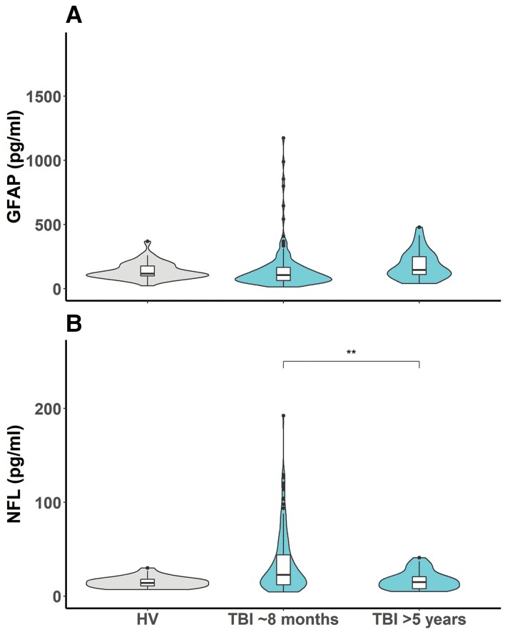
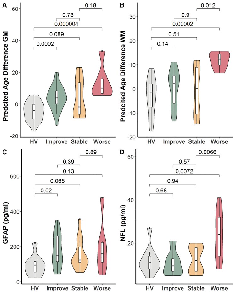
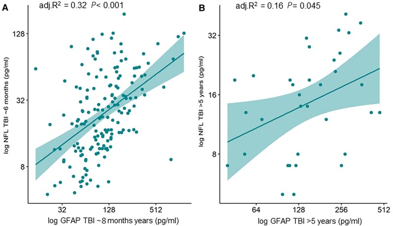
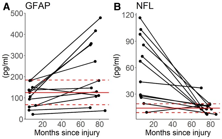
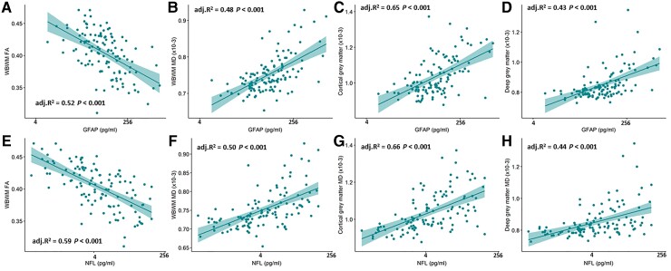
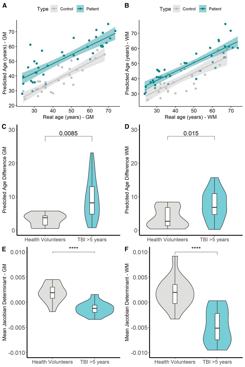
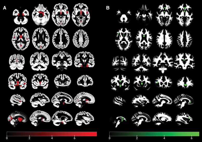
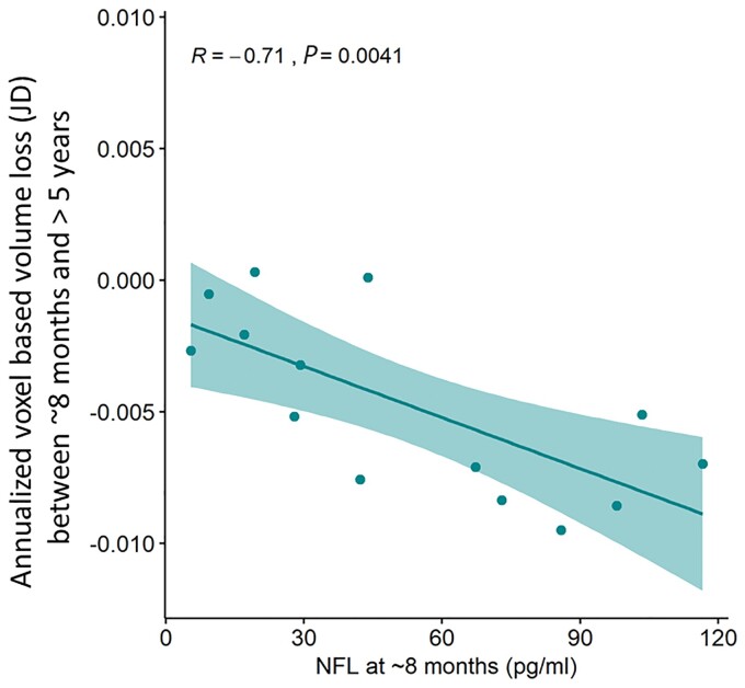
There is substantial interest in the potential for traumatic brain injury to result in progressive neurological deterioration. While blood biomarkers such as glial fibrillary acid protein (GFAP) and neurofilament light have been widely explored in characterizing acute traumatic brain injury (TBI), their use in the chronic phase is limited. Given increasing evidence that these proteins may be markers of ongoing neurodegeneration in a range of diseases, we examined their relationship to imaging changes and functional outcome in the months to years following TBI. Two-hundred and three patients were recruited in two separate cohorts; 6 months post-injury (n = 165); and >5 years post-injury (n = 38; 12 of whom also provided data ∼8 months post-TBI). Subjects underwent blood biomarker sampling (n = 199) and MRI (n = 172; including diffusion tensor imaging). Data from patient cohorts were compared to 59 healthy volunteers and 21 non-brain injury trauma controls. Mean diffusivity and fractional anisotropy were calculated in cortical grey matter, deep grey matter and whole brain white matter. Accelerated brain ageing was calculated at a whole brain level as the predicted age difference defined using T1-weighted images, and at a voxel-based level as the annualized Jacobian determinants in white matter and grey matter, referenced to a population of 652 healthy control subjects. Serum neurofilament light concentrations were elevated in the early chronic phase. While GFAP values were within the normal range at ∼8 months, many patients showed a secondary and temporally distinct elevations up to >5 years after injury. Biomarker elevation at 6 months was significantly related to metrics of microstructural injury on diffusion tensor imaging. Biomarker levels at ∼8 months predicted white matter volume loss at >5 years, and annualized brain volume loss between ∼8 months and 5 years. Patients who worsened functionally between ∼8 months and >5 years showed higher than predicted brain age and elevated neurofilament light levels. GFAP and neurofilament light levels can remain elevated months to years after TBI, and show distinct temporal profiles. These elevations correlate closely with microstructural injury in both grey and white matter on contemporaneous quantitative diffusion tensor imaging. Neurofilament light elevations at ∼8 months may predict ongoing white matter and brain volume loss over >5 years of follow-up. If confirmed, these findings suggest that blood biomarker levels at late time points could be used to identify TBI survivors who are at high risk of progressive neurological damage.
人们对创伤性脑损伤导致进行性神经恶化的潜力非常感兴趣。虽然神经丝轻链(NfL)和胶质纤维酸性蛋白(GFAP)等血液生物标志物已被广泛用于描述急性创伤性脑损伤(TBI),但其在慢性期的应用受到限制。鉴于越来越多的证据表明,这些蛋白质可能是一系列疾病中持续神经退行性变的标志物,我们研究了它们与 TBI 后数月至数年的影像学变化和功能结局的关系。在两个独立的队列中招募了 203 名患者;伤后 6 个月(n=165);以及>5 年(n=38;其中 12 人还提供了 TBI 后约 8 个月的数据)。受试者接受了血液生物标志物采样(n=199)和 MRI(n=172;包括弥散张量成像)。将患者队列的数据与 59 名健康志愿者和 21 名非颅脑外伤对照进行比较。计算皮质灰质、深部灰质和全脑白质的平均弥散度和各向异性分数。使用 T1 加权图像定义的预测年龄差异作为全脑水平的加速脑老化,以及参考 652 名健康对照受试者的白质和灰质的每年化雅可比行列式作为基于体素的水平。在早期慢性期,血清神经丝轻链浓度升高。虽然 GFAP 值在 8 个月左右仍在正常范围内,但许多患者在受伤后>5 年出现继发性和时间上明显升高。6 个月时的生物标志物升高与弥散张量成像上的微观结构损伤指标显著相关。8 个月左右的生物标志物水平预测了>5 年后的白质体积损失,以及 8 个月至 5 年间的每年脑体积损失。8 个月至>5 年期间功能恶化的患者显示出高于预测的脑龄和升高的神经丝轻链水平。TBI 后数月至数年,GFAP 和神经丝轻链水平仍升高,并呈现出不同的时间分布。这些升高与同时进行的定量弥散张量成像上的灰质和白质微观结构损伤密切相关。8 个月左右的神经丝轻链升高可能预示着>5 年的随访中持续的白质和脑体积损失。如果得到证实,这些发现表明,晚期血液生物标志物水平可用于识别 TBI 幸存者中存在进行性神经损伤风险较高的患者。