Luo Huan, Hasegawa Kayoko, Liu Mingsheng, Song Wen-Jie
Department of Sensory and Cognitive Physiology, Graduate School of Medical Sciences, Kumamoto University, Kumamoto, Japan.
Program for Leading Graduate Schools HIGO Program, Kumamoto University, Kumamoto, Japan.
Front Neuroanat. 2017 Dec 21;11:115. doi: 10.3389/fnana.2017.00115. eCollection 2017.
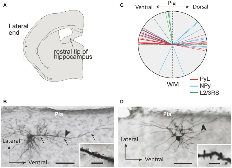
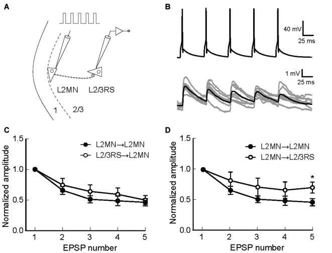
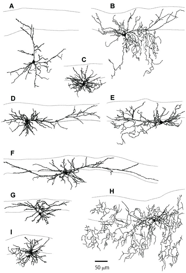
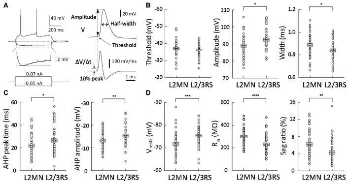
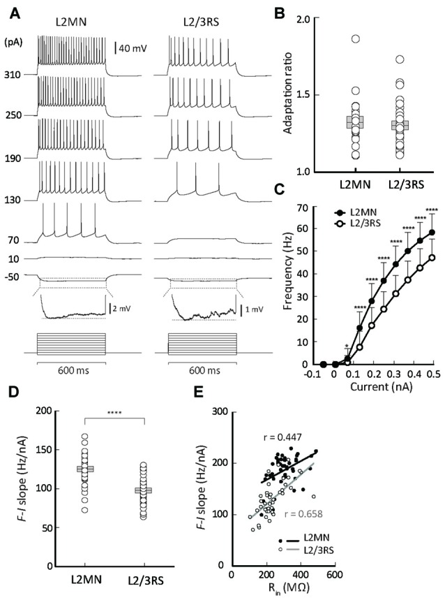
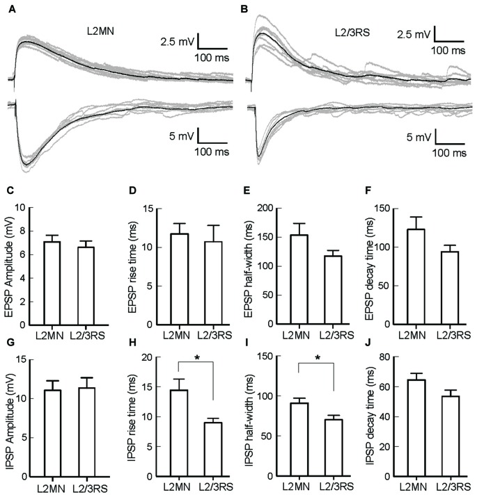
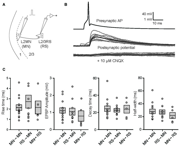
Layer 2/3 (L2/3) excitatory neurons in the neocortex make major contributions to corticocortical connections and therefore function to integrate information across cortical areas and hemispheres. Recent evidence suggests that excitatory neurons in L2/3 can have different properties. Sparse evidence from previous studies suggests that L2 neurons located at the border between L1 and L2 (referred to as L2 marginal neurons, L2MNs), have a morphology distinct from a typical pyramidal neuron. However, whether the membrane properties and input/output properties of L2MNs are different from those of typical pyramidal neurons in L2/3 is unknown. Here we addressed these questions in a slice preparation of mouse temporal cortex. We found that L2MNs were homogeneous in intrinsic membrane properties but appeared diverse in morphology. In agreement with previous studies, L2MNs either had oblique apical dendrites or had no obvious apical dendrites. The tufts of both apical and basal dendrites of these neurons invaded L1 extensively. All L2MNs showed a regular firing pattern with moderate adaptation. Compared with typical L2/3 pyramidal neurons that showed regular spiking (RS) activity (neurons), L2MNs showed a higher firing rate, larger sag ratio, and higher input resistance. No difference in the amplitude of excitatory and inhibitory postsynaptic potentials (EPSPs and IPSPs, respectively), evoked by stimulation of L1, was found between the two types of neurons, but the IPSPs in L2MNs had a slower time course than those in L2/3 RS cells. In paired recordings, unitary EPSPs showed no significant differences between synapses formed by L2MNs and those formed by L2/3 RS neurons. However, short-term synaptic depression (STSD) examined with a L2MN as the presynaptic neuron was greater when another L2MN was the postsynaptic neuron than when a L2/3 RS neuron was the postsynaptic neuron. The distinct morphological features of L2MNs found here have developmental implications, and the differences in electrophysiological properties between L2MNs and other L2/3 pyramidal neurons suggest that they play different functional roles in cortical networks.
新皮层中的第2/3层(L2/3)兴奋性神经元对皮质-皮质连接起主要作用,因此其功能是整合跨皮质区域和半球的信息。最近的证据表明,L2/3中的兴奋性神经元可能具有不同的特性。先前研究的稀少证据表明,位于L1和L2边界的L2神经元(称为L2边缘神经元,L2MNs)具有与典型锥体神经元不同的形态。然而,L2MNs的膜特性和输入/输出特性是否与L2/3中的典型锥体神经元不同尚不清楚。在这里,我们在小鼠颞叶皮质切片标本中解决了这些问题。我们发现L2MNs的内在膜特性是均匀的,但形态上似乎多种多样。与先前的研究一致,L2MNs要么有倾斜的顶树突,要么没有明显的顶树突。这些神经元的顶树突和基底树突的丛广泛侵入L1。所有L2MNs都表现出适度适应的规则放电模式。与表现出规则发放(RS)活动的典型L2/3锥体神经元相比,L2MNs表现出更高的放电率。更大的凹陷比率和更高的输入电阻。在两种类型的神经元之间,未发现刺激L1诱发的兴奋性和抑制性突触后电位(分别为EPSPs和IPSPs)幅度有差异,但L2MNs中的IPSPs时程比L2/3 RS细胞中的慢。在配对记录中,由L2MNs形成的突触和由L2/3 RS神经元形成的突触之间,单一EPSPs没有显著差异。然而,当以另一个L2MN为突触后神经元时,以L2MN为突触前神经元检测到的短期突触抑制(STSD)比以L2/3 RS神经元为突触后神经元时更大。这里发现的L2MNs独特的形态特征具有发育意义,L2MNs与其他L2/3锥体神经元在电生理特性上的差异表明它们在皮质网络中发挥不同的功能作用。