Albi Angela, Meola Antonio, Zhang Fan, Kahali Pegah, Rigolo Laura, Tax Chantal M W, Ciris Pelin Aksit, Essayed Walid I, Unadkat Prashin, Norton Isaiah, Rathi Yogesh, Olubiyi Olutayo, Golby Alexandra J, O'Donnell Lauren J
Brigham and Women's Hospital, Harvard Medical School, Boston, MA.
Center for Mind/Brain Sciences (CIMEC), University of Trento, Rovereto, Italy.
J Neuroimaging. 2018 Mar;28(2):173-182. doi: 10.1111/jon.12485. Epub 2018 Jan 10.
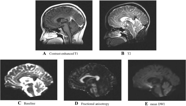
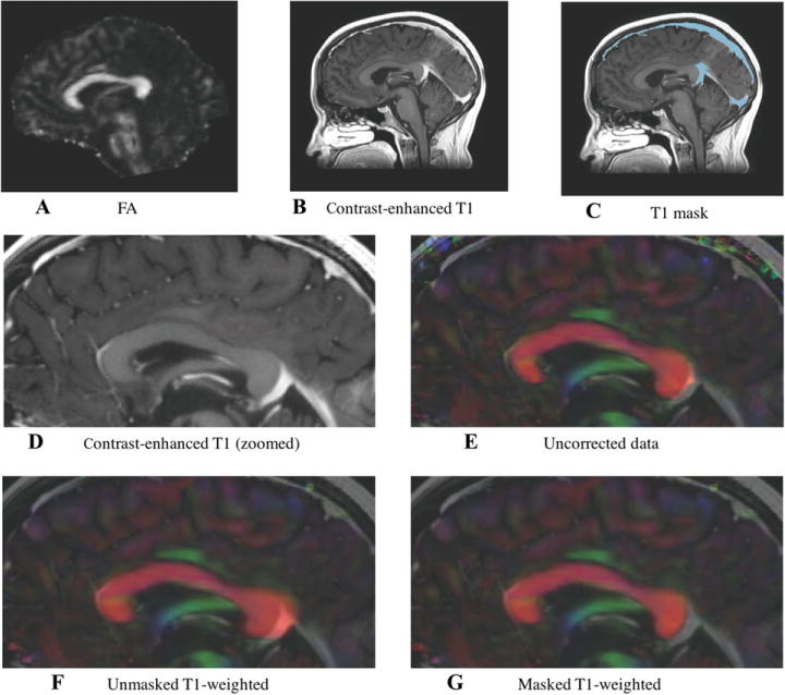
Diffusion magnetic resonance imaging (dMRI) provides preoperative maps of neurosurgical patients' white matter tracts, but these maps suffer from echo-planar imaging (EPI) distortions caused by magnetic field inhomogeneities. In clinical neurosurgical planning, these distortions are generally not corrected and thus contribute to the uncertainty of fiber tracking. Multiple image processing pipelines have been proposed for image-registration-based EPI distortion correction in healthy subjects. In this article, we perform the first comparison of such pipelines in neurosurgical patient data.
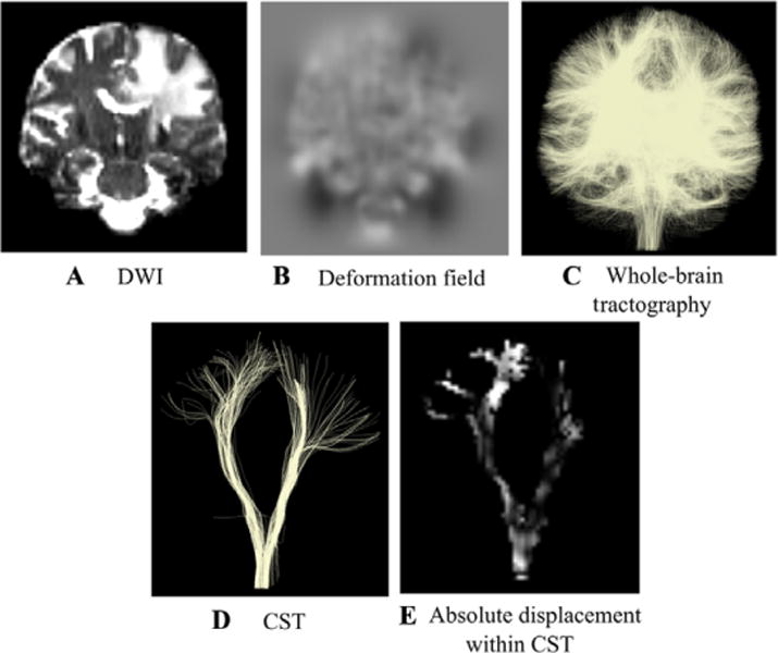
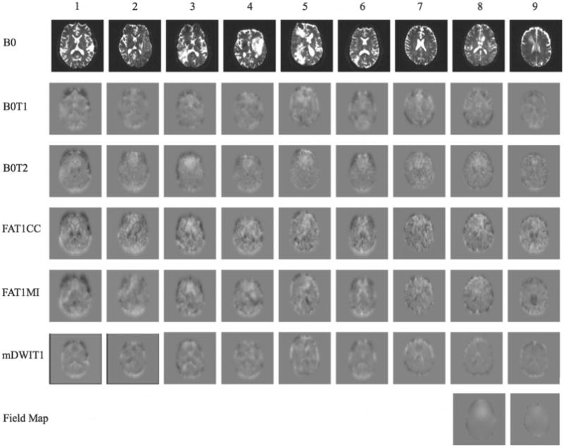
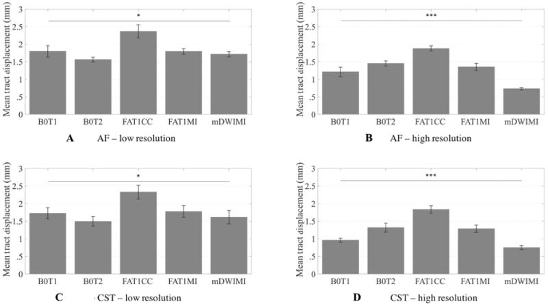
Five pipelines were tested in a retrospective clinical dMRI dataset of 9 patients with brain tumors. Pipelines differed in the choice of fixed and moving images and the similarity metric for image registration. Distortions were measured in two important tracts for neurosurgery, the arcuate fasciculus and corticospinal tracts.
Significant differences in distortion estimates were found across processing pipelines. The most successful pipeline used dMRI baseline and T2-weighted images as inputs for distortion correction. This pipeline gave the most consistent distortion estimates across image resolutions and brain hemispheres.
Quantitative results of mean tract distortions on the order of 1-2 mm are in line with other recent studies, supporting the potential need for distortion correction in neurosurgical planning. Novel results include significantly higher distortion estimates in the tumor hemisphere and greater effect of image resolution choice on results in the tumor hemisphere. Overall, this study demonstrates possible pitfalls and indicates that care should be taken when implementing EPI distortion correction in clinical settings.
扩散磁共振成像(dMRI)可为神经外科手术患者提供白质束的术前图谱,但这些图谱会受到磁场不均匀性导致的回波平面成像(EPI)畸变的影响。在临床神经外科手术规划中,这些畸变通常未得到校正,从而增加了纤维追踪的不确定性。针对健康受试者,已经提出了多种基于图像配准的EPI畸变校正的图像处理流程。在本文中,我们首次在神经外科患者数据中对这些流程进行了比较。
在一个包含9例脑肿瘤患者的回顾性临床dMRI数据集中测试了五种流程。这些流程在固定图像和移动图像的选择以及图像配准的相似性度量方面存在差异。在神经外科手术的两个重要束,即弓状束和皮质脊髓束中测量畸变。
在不同的处理流程中发现了畸变估计的显著差异。最成功的流程使用dMRI基线图像和T2加权图像作为畸变校正的输入。该流程在不同图像分辨率和脑半球上给出了最一致的畸变估计。
平均束畸变在1 - 2毫米量级的定量结果与其他近期研究一致,支持了神经外科手术规划中可能需要进行畸变校正的观点。新的结果包括肿瘤半球的畸变估计显著更高,以及图像分辨率选择对肿瘤半球结果的影响更大。总体而言,本研究展示了可能存在的问题,并表明在临床环境中实施EPI畸变校正时应谨慎。