• 文献检索
  • 文档翻译
  • 深度研究
  • 学术资讯
  • Suppr Zotero 插件Zotero 插件
  • 邀请有礼
  • 套餐&价格
  • 历史记录
应用&插件
Suppr Zotero 插件Zotero 插件浏览器插件Mac 客户端Windows 客户端微信小程序
定价
高级版会员购买积分包购买API积分包
服务
文献检索文档翻译深度研究API 文档MCP 服务
关于我们
关于 Suppr公司介绍联系我们用户协议隐私条款
关注我们

Suppr 超能文献

核心技术专利:CN118964589B侵权必究
粤ICP备2023148730 号-1Suppr @ 2026

文献检索

告别复杂PubMed语法,用中文像聊天一样搜索,搜遍4000万医学文献。AI智能推荐,让科研检索更轻松。

立即免费搜索

文件翻译

保留排版,准确专业,支持PDF/Word/PPT等文件格式,支持 12+语言互译。

免费翻译文档

深度研究

AI帮你快速写综述,25分钟生成高质量综述,智能提取关键信息,辅助科研写作。

立即免费体验

大鼠腹膜肥大细胞暴露于矿物质纤维后的分泌反应。

The Secretory Response of Rat Peritoneal Mast Cells on Exposure to Mineral Fibers.

机构信息

Department of Life Science University of Trieste, University of Trieste, 34127 Trieste, Italy.

出版信息

Int J Environ Res Public Health. 2018 Jan 10;15(1):104. doi: 10.3390/ijerph15010104.

DOI:10.3390/ijerph15010104
PMID:29320402
原文链接:https://pmc.ncbi.nlm.nih.gov/articles/PMC5800203/
Abstract

BACKGROUND

Exposure to mineral fibers is of substantial relevance to human health. A key event in exposure is the interaction with inflammatory cells and the subsequent generation of pro-inflammatory factors. Mast cells (MCs) have been shown to interact with titanium oxide (TiO₂) and asbestos fibers. In this study, we compared the response of rat peritoneal MCs challenged with the asbestos crocidolite and nanowires of TiO₂ to that induced by wollastonite employed as a control fiber.

METHODS

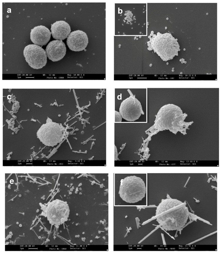
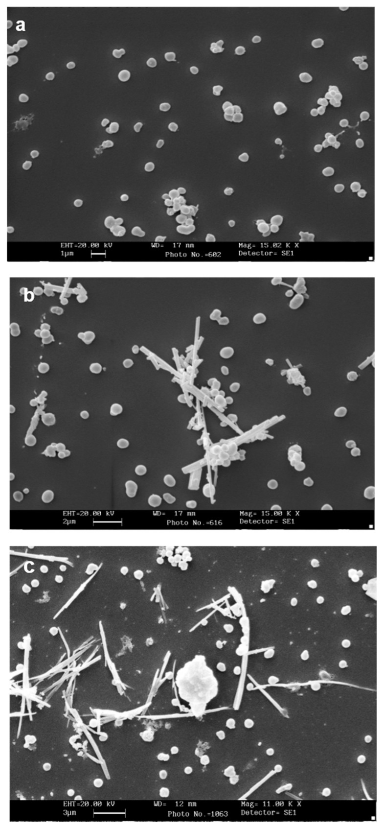
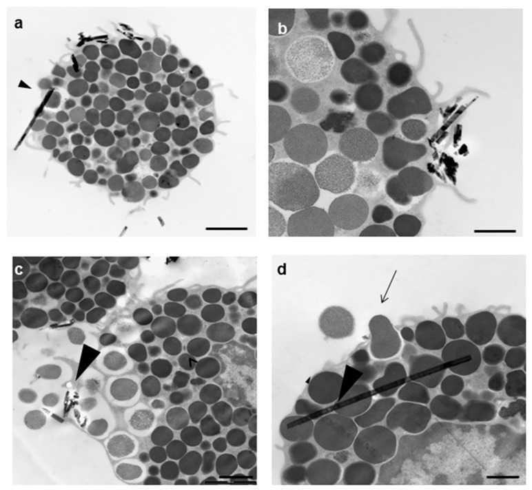
Rat peritoneal MCs (RPMCs), isolated from peritoneal lavage, were incubated in the presence of mineral fibers. The quantities of secreted enzymes were evaluated together with the activity of fiber-associated enzymes. The ultrastructural morphology of fiber-interacting RPMCs was analyzed with electron microscopy.

RESULTS

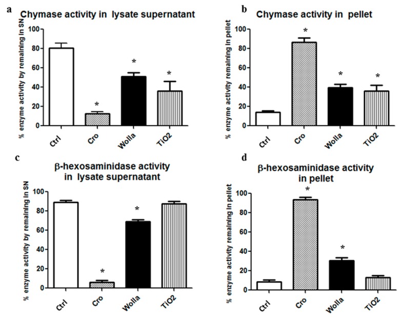
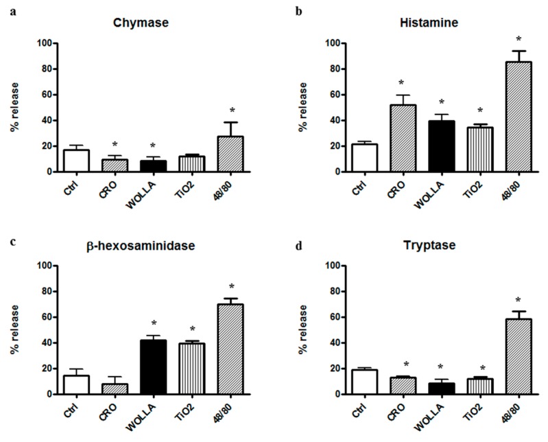
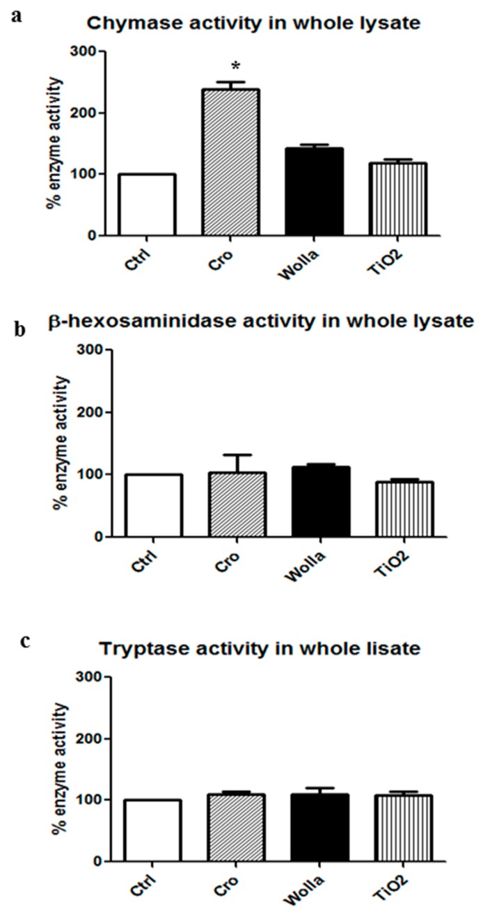
Asbestos and TiO₂ stimulate MC secretion. Secreted enzymes bind to fibers and exhibit higher activity. TiO₂ and wollastonite bind and improve enzyme activity, but to a lesser degree than crocidolite.

CONCLUSIONS

(1) Mineral fibers are able to stimulate the mast cell secretory process by both active (during membrane interaction) and/or passive (during membrane penetration) interaction; (2) fibers can be found to be associated with secreted enzymes-this process appears to create long-lasting pro-inflammatory environments and may represent the active contribution of MCs in maintaining the inflammatory process; (3) MCs and their enzymes should be considered as a therapeutic target in the pathogenesis of asbestos-induced lung inflammation; and (4) MCs can contribute to the inflammatory effect associated with selected engineered nanomaterials, such as TiO₂ nanoparticles.

摘要

背景

暴露于矿物质纤维对人类健康具有重要意义。暴露的一个关键事件是与炎症细胞相互作用,以及随后产生促炎因子。已经表明肥大细胞(MC)与二氧化钛(TiO₂)和石棉纤维相互作用。在这项研究中,我们比较了大鼠腹膜肥大细胞(RPMC)与石棉青石棉和 TiO₂纳米线挑战,以及作为对照纤维的硅灰石诱导的反应。

方法

从腹膜灌洗中分离大鼠腹膜肥大细胞(RPMC),在存在矿物质纤维的情况下孵育。评估分泌酶的量以及纤维相关酶的活性。用电子显微镜分析与纤维相互作用的 RPMC 的超微结构形态。

结果

石棉和 TiO₂刺激 MC 分泌。分泌的酶与纤维结合并表现出更高的活性。TiO₂和硅灰石结合并提高了酶的活性,但程度低于青石棉。

结论

(1)矿物质纤维能够通过主动(在膜相互作用期间)和/或被动(在膜穿透期间)相互作用刺激肥大细胞分泌过程; (2)纤维可以与分泌的酶相关联-这个过程似乎创造了持久的促炎环境,并可能代表 MC 维持炎症过程的积极贡献; (3)MC 及其酶应被视为石棉诱导的肺炎症发病机制中的治疗靶标; (4)MC 可导致与选定的工程纳米材料(如 TiO₂纳米粒子)相关的炎症效应。

https://cdn.ncbi.nlm.nih.gov/pmc/blobs/49bc/5800203/f1ae14e73380/ijerph-15-00104-g008.jpg
https://cdn.ncbi.nlm.nih.gov/pmc/blobs/49bc/5800203/0dbc52975130/ijerph-15-00104-g001a.jpg
https://cdn.ncbi.nlm.nih.gov/pmc/blobs/49bc/5800203/4f403ee894d0/ijerph-15-00104-g002.jpg
https://cdn.ncbi.nlm.nih.gov/pmc/blobs/49bc/5800203/493136f89e5a/ijerph-15-00104-g003.jpg
https://cdn.ncbi.nlm.nih.gov/pmc/blobs/49bc/5800203/4d17869c4783/ijerph-15-00104-g004.jpg
https://cdn.ncbi.nlm.nih.gov/pmc/blobs/49bc/5800203/441494d96c63/ijerph-15-00104-g005.jpg
https://cdn.ncbi.nlm.nih.gov/pmc/blobs/49bc/5800203/edf5feb4c5fb/ijerph-15-00104-g006.jpg
https://cdn.ncbi.nlm.nih.gov/pmc/blobs/49bc/5800203/ce642b3478ba/ijerph-15-00104-g007.jpg
https://cdn.ncbi.nlm.nih.gov/pmc/blobs/49bc/5800203/f1ae14e73380/ijerph-15-00104-g008.jpg

相似文献

1
The Secretory Response of Rat Peritoneal Mast Cells on Exposure to Mineral Fibers.大鼠腹膜肥大细胞暴露于矿物质纤维后的分泌反应。
Int J Environ Res Public Health. 2018 Jan 10;15(1):104. doi: 10.3390/ijerph15010104.
2
Mesothelial cell proliferation and biopersistence of wollastonite and crocidolite asbestos fibers.间皮细胞增殖以及硅灰石和青石棉纤维的生物持久性
Fundam Appl Toxicol. 1997 Aug;38(2):173-83. doi: 10.1006/faat.1997.2344.
3
Characterization of pulmonary responses in mice to asbestos/asbestiform fibers using gene expression profiles.利用基因表达谱对小鼠肺部对石棉/似石棉纤维的反应进行特征描述。
J Toxicol Environ Health A. 2018;81(4):60-79. doi: 10.1080/15287394.2017.1408201. Epub 2017 Dec 26.
4
Wollastonite fibers in vitro generate reactive oxygen species able to lyse erythrocytes and activate the complement alternate pathway.硅灰石纤维在体外可产生活性氧,这些活性氧能够裂解红细胞并激活补体替代途径。
Toxicol Sci. 1998 Jul;44(1):32-8. doi: 10.1006/toxs.1998.2440.
5
Differential in vitro cellular response induced by exposure to synthetic vitreous fibers (SVFs) and asbestos crocidolite fibers.暴露于合成玻璃纤维(SVF)和温石棉纤维所诱导的体外细胞反应差异。
Exp Mol Pathol. 2006 Aug;81(1):31-41. doi: 10.1016/j.yexmp.2005.10.001. Epub 2005 Dec 13.
6
Combined effect of selected mineral fibres and tobacco smoke on respiratory tract in rats.特定矿物纤维与烟草烟雾对大鼠呼吸道的联合作用
Cent Eur J Public Health. 2014 Sep;22(3):159-63. doi: 10.21101/cejph.a3894.
7
Asbestos induces apoptosis in human alveolar macrophages.石棉可诱导人肺泡巨噬细胞凋亡。
Am J Physiol. 1996 Nov;271(5 Pt 1):L813-9. doi: 10.1152/ajplung.1996.271.5.L813.
8
Macrophage phagocytic activity and cell phagocytic capacity following intratracheal instillation of a comixture of asbestos fibers.气管内滴注石棉纤维混合物后巨噬细胞的吞噬活性和细胞吞噬能力
Exp Toxicol Pathol. 1995 Sep;47(4):238-40. doi: 10.1016/S0940-2993(11)80256-3.
9
Pleural macrophage recruitment and activation in asbestos-induced pleural injury.石棉诱导的胸膜损伤中胸膜巨噬细胞的募集与激活
Environ Health Perspect. 1997 Sep;105 Suppl 5(Suppl 5):1257-60. doi: 10.1289/ehp.97105s51257.
10
Tumorigenicity of cellulose fibers injected into the rat peritoneal cavity.注入大鼠腹腔的纤维素纤维的致瘤性。
Inhal Toxicol. 2002 Jul;14(7):685-703. doi: 10.1080/08958370290084584.

引用本文的文献

1
Ferritin adsorption onto chrysotile asbestos fibers influences the protein secondary structure.铁蛋白吸附到温石棉纤维上会影响蛋白质的二级结构。
Heliyon. 2024 Oct 4;10(20):e38966. doi: 10.1016/j.heliyon.2024.e38966. eCollection 2024 Oct 30.
2
Asbestos Fibers Enhance the TMEM16A Channel Activity in Xenopus Oocytes.石棉纤维增强非洲爪蟾卵母细胞中的TMEM16A通道活性。
Membranes (Basel). 2023 Feb 1;13(2):180. doi: 10.3390/membranes13020180.
3
Autoantibodies and cancer among asbestos-exposed cohorts in Western Australia.澳大利亚西部暴露于石棉人群中的自身抗体与癌症。

本文引用的文献

1
Mast cell chymase promotes hypertrophic scar fibroblast proliferation and collagen synthesis by activating TGF-β1/Smads signaling pathway.肥大细胞糜蛋白酶通过激活TGF-β1/Smads信号通路促进增生性瘢痕成纤维细胞增殖和胶原蛋白合成。
Exp Ther Med. 2017 Nov;14(5):4438-4442. doi: 10.3892/etm.2017.5082. Epub 2017 Aug 31.
2
Titanium dioxide nanoparticles prime a specific activation state of macrophages.二氧化钛纳米颗粒引发巨噬细胞的特定激活状态。
Nanotoxicology. 2017 Aug;11(6):737-750. doi: 10.1080/17435390.2017.1349202. Epub 2017 Jul 15.
3
Impact of anatase titanium dioxide nanoparticles on mutagenic and genotoxic response in Chinese hamster lung fibroblast cells (V-79): The role of cellular uptake.
J Toxicol Environ Health A. 2021 Jun 3;84(11):475-483. doi: 10.1080/15287394.2021.1889424. Epub 2021 Mar 7.
4
The Effects of Asbestos Fibers on Human T Cells.石棉纤维对人 T 细胞的影响。
Int J Mol Sci. 2020 Sep 23;21(19):6987. doi: 10.3390/ijms21196987.
5
Mast Cells in Peritoneal Fluid From Women With Endometriosis and Their Possible Role in Modulating Sperm Function.子宫内膜异位症女性腹腔液中的肥大细胞及其在调节精子功能中的可能作用。
Front Physiol. 2020 Jan 9;10:1543. doi: 10.3389/fphys.2019.01543. eCollection 2019.
6
On the mechanism of the electrophysiological changes and membrane lesions induced by asbestos fiber exposure in Xenopus laevis oocytes.探讨石棉纤维暴露致非洲爪蟾卵母细胞电生理改变及膜损伤的机制。
Sci Rep. 2019 Feb 14;9(1):2014. doi: 10.1038/s41598-019-38591-x.
锐钛矿型二氧化钛纳米颗粒对中国仓鼠肺成纤维细胞(V-79)诱变和遗传毒性反应的影响:细胞摄取的作用。
Food Chem Toxicol. 2017 Jul;105:127-139. doi: 10.1016/j.fct.2017.04.005. Epub 2017 Apr 8.
4
The Effect of Ambient Titanium Dioxide Microparticle Exposure to the Ocular Surface on the Expression of Inflammatory Cytokines in the Eye and Cervical Lymph Nodes.环境中二氧化钛微粒暴露于眼表对眼部及颈部淋巴结中炎性细胞因子表达的影响
Invest Ophthalmol Vis Sci. 2016 Dec 1;57(15):6580-6590. doi: 10.1167/iovs.16-19944.
5
Markers of lipid oxidative damage in the exhaled breath condensate of nano TiO production workers.纳米二氧化钛生产工人呼出气冷凝物中脂质氧化损伤的标志物。
Nanotoxicology. 2017 Feb;11(1):52-63. doi: 10.1080/17435390.2016.1262921. Epub 2016 Dec 9.
6
Leukotrienes in exhaled breath condensate and fractional exhaled nitric oxide in workers exposed to TiO2 nanoparticles.暴露于二氧化钛纳米颗粒的工人呼出气体冷凝物中的白三烯和呼出一氧化氮分数
J Breath Res. 2016 Jun 30;10(3):036004. doi: 10.1088/1752-7155/10/3/036004.
7
Role of Macrophage (M1 and M2) in Titanium-Dioxide Nanoparticle-Induced Oxidative Stress and Inflammatory Response in Rat.巨噬细胞(M1和M2)在二氧化钛纳米颗粒诱导的大鼠氧化应激和炎症反应中的作用
Appl Biochem Biotechnol. 2016 Dec;180(7):1257-1275. doi: 10.1007/s12010-016-2165-x. Epub 2016 Jun 18.
8
The Third Italian Consensus Conference for Malignant Pleural Mesothelioma: State of the art and recommendations.第三届意大利恶性胸膜间皮瘤共识会议:现状与建议。
Crit Rev Oncol Hematol. 2016 Aug;104:9-20. doi: 10.1016/j.critrevonc.2016.05.004. Epub 2016 May 13.
9
The proliferative effects of asbestos-exposed peripheral blood mononuclear cells on mesothelial cells.石棉暴露的外周血单个核细胞对间皮细胞的增殖作用。
Oncol Lett. 2016 May;11(5):3308-3316. doi: 10.3892/ol.2016.4412. Epub 2016 Apr 5.
10
Proteomic changes in human lung epithelial cells (A549) in response to carbon black and titanium dioxide exposures.人类肺上皮细胞(A549)暴露于炭黑和二氧化钛后的蛋白质组变化。
J Proteomics. 2016 Oct 21;149:53-63. doi: 10.1016/j.jprot.2016.03.046. Epub 2016 Apr 12.