Department of Biology, Brandon University, Brandon, Manitoba, Canada.
Department of Anatomy and Cell Biology, Schulich School of Medicine and Dentistry, University of Western Ontario, London, Ontario, Canada.
Sci Rep. 2018 Jan 10;8(1):327. doi: 10.1038/s41598-017-18612-3.
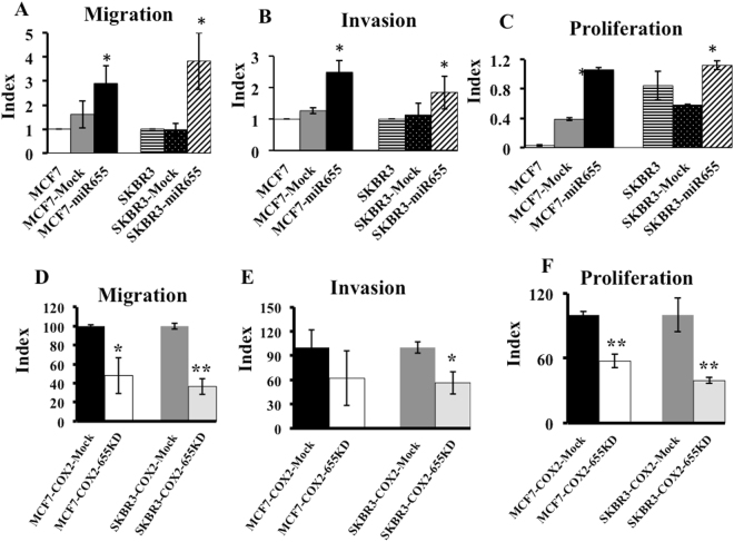
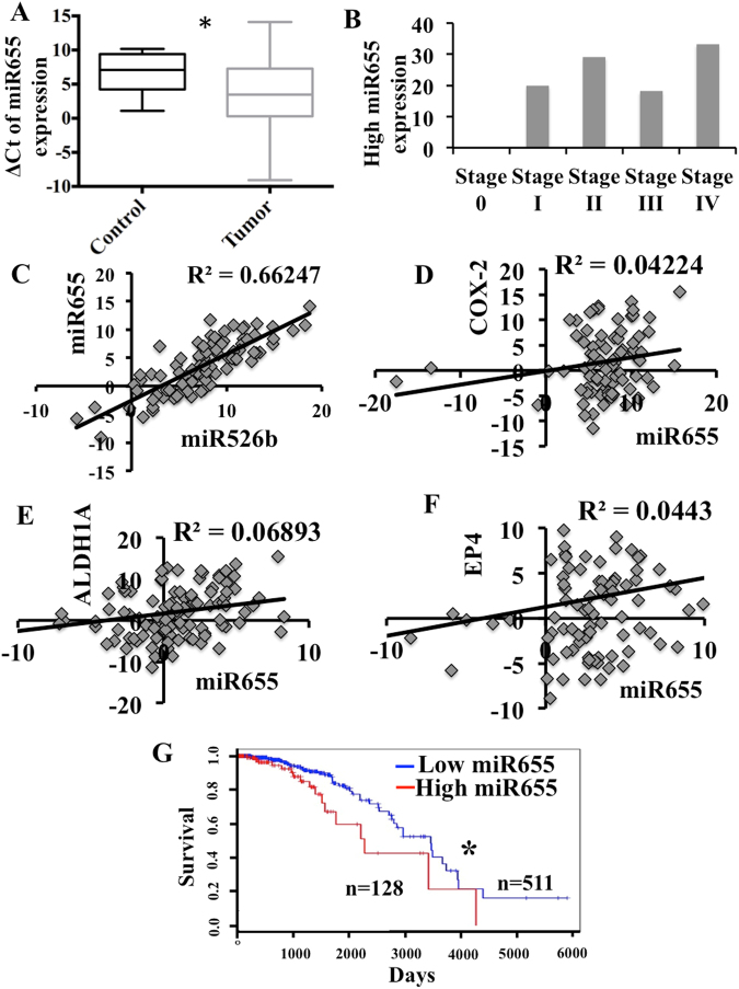
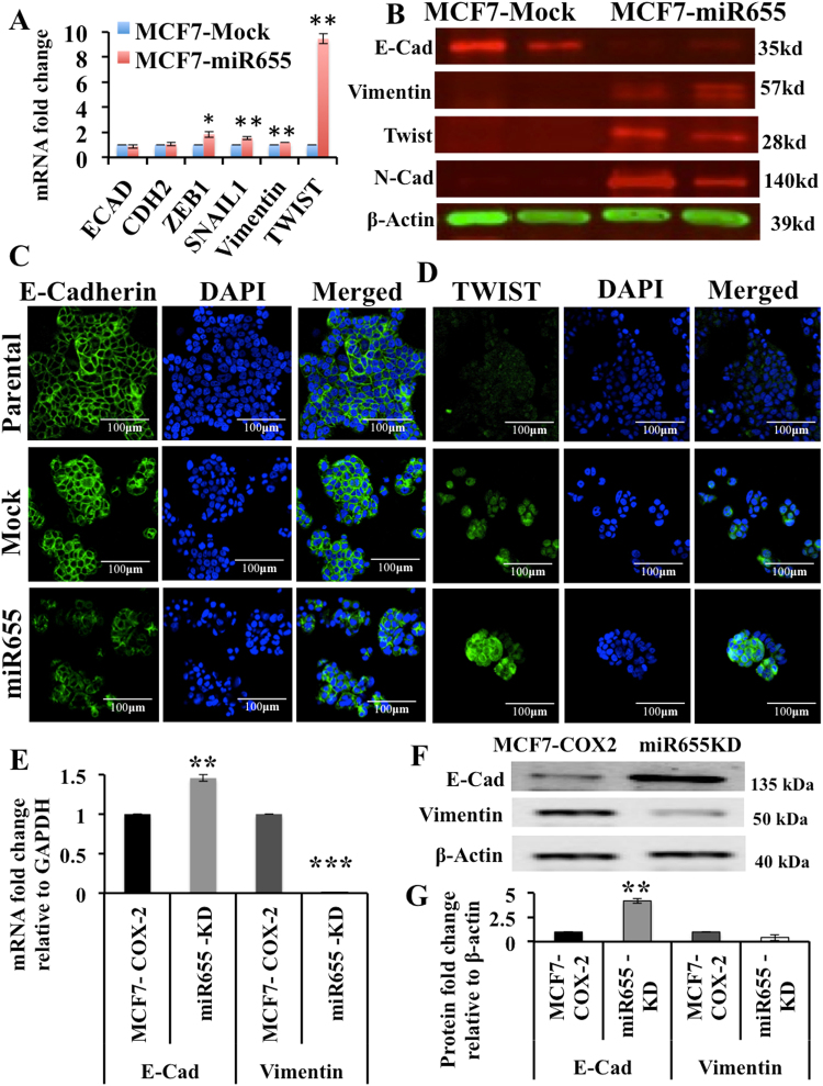
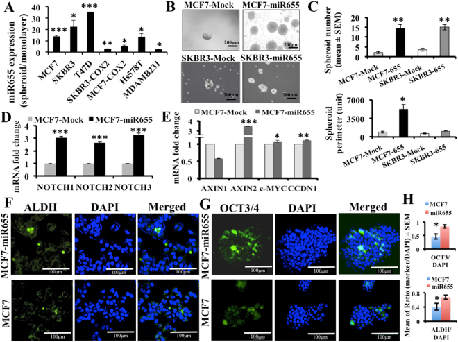
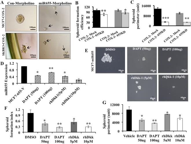
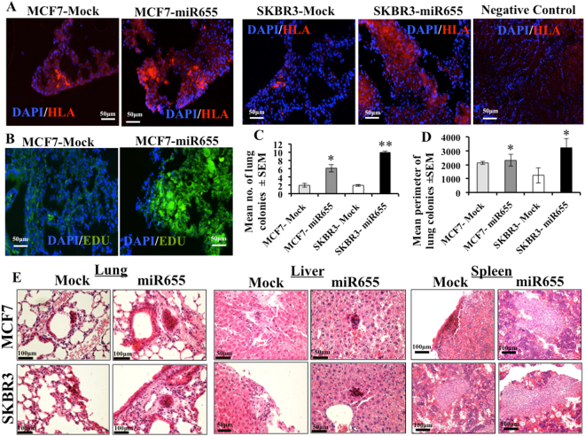
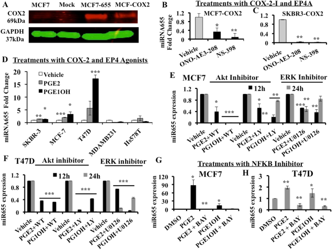
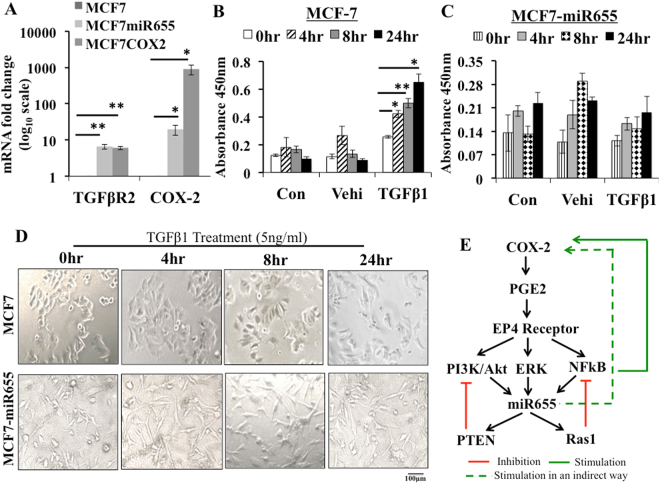
We show that Cyclooxygenase-2 over-expression induces an oncogenic microRNA miR655 in human breast cancer cells by activation of EP4. MiR655 expression positively correlated with COX-2 in genetically disparate breast cancer cell lines and increased in all cell lines when grown as spheroids, implicating its link with stem-like cells (SLCs). Ectopic miR655 over-expression in MCF7 and SKBR3 cells resulted in increased proliferation, migration, invasion, spheroid formation and Epithelial to Masenchymal transition (EMT). Conversely, knocking down miR655 in aggressive MCF7-COX2 and SKBR3-COX2 cells reverted these phenotypes. MCF7-miR655 cells displayed upregulated NOTCH/WNT genes; both pathway inhibitors abrogated miR655-induced spheroid formation, linking miR655 with SLC-related pathways. MiR655 expression was dependent on EP4 activity and EP4 downstream signaling pathways PI3K/AKT, ERK and NF-kB and led to TGFβ resistance for Smad3 phosphorylation. Tail vein injection of MCF7-miR655 and SKBR3-miR655 cells in NOD/SCID/GUSB-null mice revealed increased lung colony growth and micrometastases to liver and spleen. MiR655 expression was significantly high in human breast tumors (n = 105) compared to non-tumor tissues (n = 20) and associated with reduced patient survival. Thus miR655 could serve as a prognostic breast cancer biomarker.
我们表明,环氧合酶-2(COX-2)过表达通过激活 EP4 诱导人类乳腺癌细胞中的致癌 microRNA miR655。miR655 的表达与遗传上不同的乳腺癌细胞系中的 COX-2 呈正相关,并且在所有细胞系中以球体形式生长时都会增加,暗示其与干细胞样细胞(SLCs)有关。MCF7 和 SKBR3 细胞中外源性 miR655 的过表达导致增殖、迁移、侵袭、球体形成和上皮间质转化(EMT)增加。相反,在侵袭性 MCF7-COX2 和 SKBR3-COX2 细胞中敲低 miR655 则使这些表型逆转。MCF7-miR655 细胞显示出 NOTCH/WNT 基因的上调;两种途径抑制剂均消除了 miR655 诱导的球体形成,将 miR655 与 SLC 相关途径联系起来。miR655 的表达依赖于 EP4 活性及其下游信号通路 PI3K/AKT、ERK 和 NF-kB,并导致 TGFβ 对 Smad3 磷酸化的抗性。MCF7-miR655 和 SKBR3-miR655 细胞在 NOD/SCID/GUSB-null 小鼠的尾静脉注射显示出肺部集落生长增加和向肝和脾的微转移。与非肿瘤组织(n=20)相比,miR655 在人类乳腺癌肿瘤(n=105)中的表达显著升高,与患者生存时间减少相关。因此,miR655 可作为一种预后性乳腺癌生物标志物。