Department of Radiation Oncology, University Hospital of Heidelberg, Heidelberg, Germany.
Department of Medical Physics in Radiation Oncology (E040), German Cancer Research Center (DKFZ), Im Neuenheimer Feld 280, 69120, Heidelberg, Germany.
Radiat Oncol. 2018 Jan 11;13(1):5. doi: 10.1186/s13014-017-0950-5.
The present work summarizes the research activities on radiation-induced late effects in the rat spinal cord carried out within the "clinical research group ion beam therapy" funded by the German Research Foundation (DFG, KFO 214).
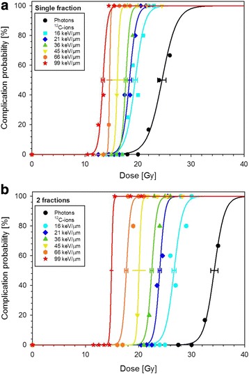
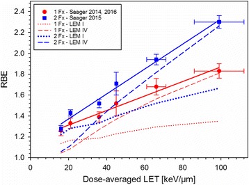
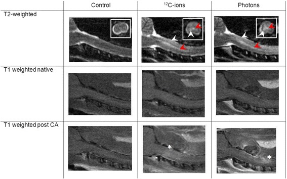
Dose-response curves for the endpoint radiation-induced myelopathy were determined at 6 different positions (LET 16-99 keV/μm) within a 6 cm spread-out Bragg peak using either 1, 2 or 6 fractions of carbon ions. Based on the tolerance dose TD of carbon ions and photons, the relative biological effectiveness (RBE) was determined and compared with predictions of the local effect model (LEM I and IV). Within a longitudinal magnetic resonance imaging (MRI)-based study the temporal development of radiation-induced changes in the spinal cord was characterized. To test the protective potential of the ACE (angiotensin converting enzyme)-inhibitor ramipril™, an additional dose-response experiment was performed.
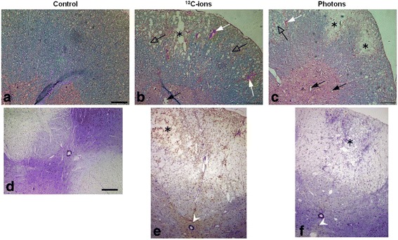
The RBE-values increased with LET and the increase was found to be larger for smaller fractional doses. Benchmarking the RBE-values as predicted by LEM I and LEM IV with the measured data revealed that LEM IV is more accurate in the high-LET, while LEM I is more accurate in the low-LET region. Characterization of the temporal development of radiation-induced changes with MRI demonstrated a shorter latency time for carbon ions, reflected on the histological level by an increased vessel perforation after carbon ion as compared to photon irradiations. For the ACE-inhibitor ramipril™, a mitigative rather than protective effect was found.
This comprehensive study established a large and consistent RBE data base for late effects in the rat spinal cord after carbon ion irradiation which will be further extended in ongoing studies. Using MRI, an extensive characterization of the temporal development of radiation-induced alterations was obtained. The reduced latency time for carbon ions is expected to originate from a dynamic interaction of various complex pathological processes. A dominant observation after carbon ion irradiation was an increase in vessel perforation preferentially in the white matter. To enable a targeted pharmacological intervention more details of the molecular pathways, responsible for the development of radiation-induced myelopathy are required.
本工作总结了德国研究基金会(DFG,KFO 214)资助的“临床研究组离子束治疗”中进行的大鼠脊髓辐射诱导晚期效应研究活动。
在 6 厘米扩展布拉格峰内,使用 1、2 或 6 个碳离子分数,在 6 个不同位置(LET 16-99keV/μm)确定了终点辐射诱导脊髓病的剂量反应曲线。基于碳离子和光子的耐受剂量 TD,确定了相对生物学效应(RBE),并与局部效应模型(LEM I 和 IV)的预测进行了比较。在基于纵向磁共振成像(MRI)的研究中,表征了脊髓中辐射诱导变化的时间发展。为了测试 ACE(血管紧张素转换酶)抑制剂雷米普利™的保护潜力,进行了额外的剂量反应实验。
RBE 值随 LET 增加而增加,并且对于较小的分数剂量,增加幅度更大。将 LEM I 和 LEM IV 预测的 RBE 值与测量数据进行基准测试表明,LEM IV 在高 LET 下更准确,而 LEM I 在低 LET 区域更准确。使用 MRI 对辐射诱导变化的时间发展进行特征描述表明,碳离子的潜伏期更短,在组织学水平上,与光子照射相比,碳离子照射后血管穿孔增加。对于 ACE 抑制剂雷米普利™,发现其具有缓解作用而非保护作用。
这项全面的研究为碳离子照射后大鼠脊髓晚期效应建立了一个大型且一致的 RBE 数据库,该数据库将在正在进行的研究中进一步扩展。使用 MRI,获得了对辐射诱导改变的时间发展的广泛特征描述。碳离子潜伏期缩短预计源于各种复杂病理过程的动态相互作用。碳离子照射后一个主要观察结果是血管穿孔增加,主要发生在白质中。为了实现靶向药物干预,需要更多关于导致辐射诱导脊髓病的分子途径的详细信息。