Roche-Campo Ferran, Bedet Alexandre, Vivier Emmanuel, Brochard Laurent, Mekontso Dessap Armand
Service de Réanimation Médicale, DHU A-TVB, Hôpitaux Universitaires Henri Mondor, Assistance Publique - Hôpitaux de Paris, 51 Avenue du Maréchal de Lattre de Tassigny, 94010, Créteil Cedex, France.
Servei de Medicina Intensiva, Hospital Verge de la Cinta, Tortosa, Tarragona, Spain.
Ann Intensive Care. 2018 Jan 9;8(1):2. doi: 10.1186/s13613-017-0348-4.
Cardiac dysfunction is a common cause of weaning failure. Weaning shares some similarities with a cardiac stress test and may challenge active phases of the cardiac cycle-like ventricular contractility and relaxation. This study aimed at assessing systolic and diastolic function during the weaning process and scrutinizing their dynamics during weaning trials.
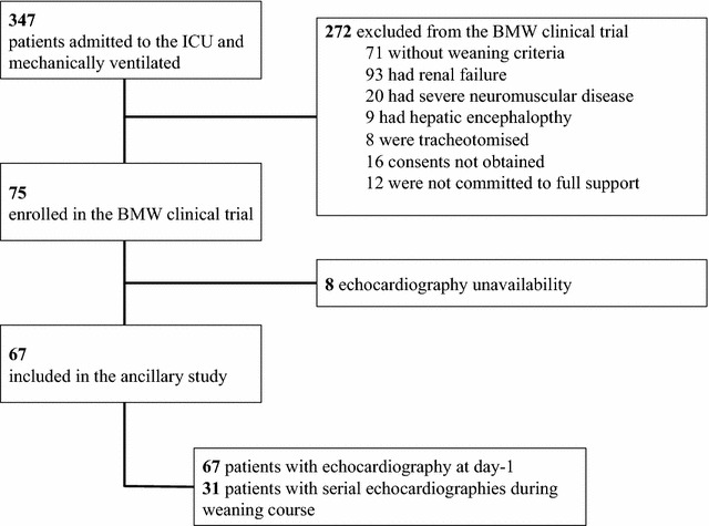
Echocardiography was performed during baseline ventilator settings to assess cardiac function at the initiation of the weaning process and at the start and the end of consecutive weaning trials (performed at day-1, day-2, and before extubation if applicable) to explore the evolution of left ventricle contractility and relaxation in a subset of patients.
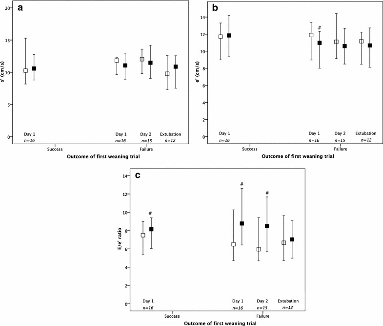
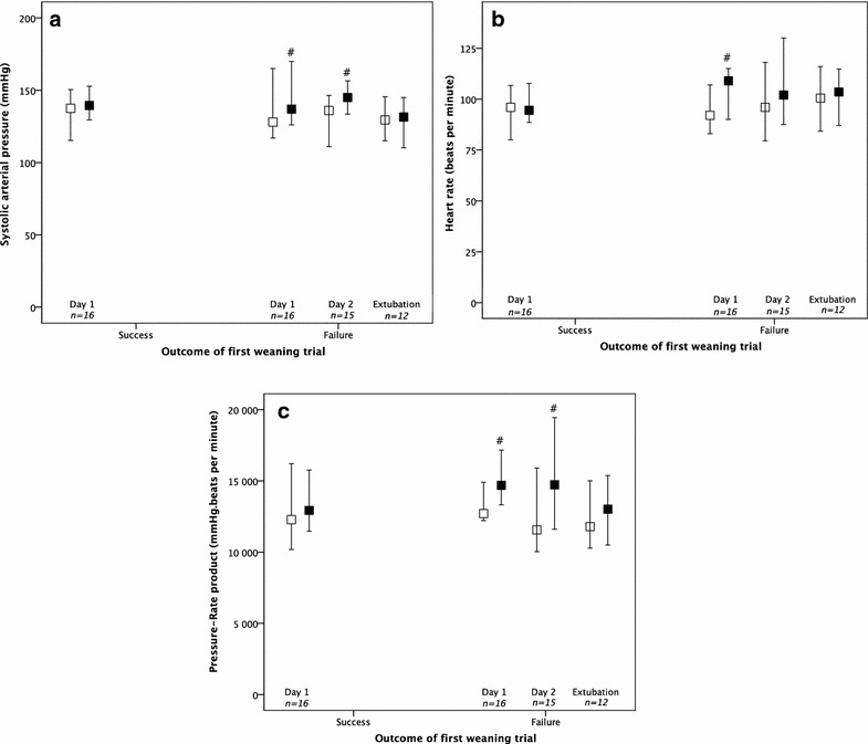
Among 67 patients included, weaning was prolonged (≥ 7 days) in 18 (27%) patients and short (< 7 days) in 49 (73%). Prevalence of systolic dysfunction and isolated diastolic dysfunction before the initiation of weaning process were 37 and 17%, respectively. Isolated diastolic dysfunction was more frequent in patients with prolonged weaning as compared to their counterparts. Thirty-one patients were explored by echocardiography during consecutive weaning trials. An increase in filling pressures with an alteration of ventricular relaxation (as assessed by a decrease in tissue Doppler early mitral diastolic wave velocity) was found during failed weaning trials.
Isolated diastolic dysfunction was associated with a prolongation of weaning. Increased filling pressures with left ventricle relaxation impairment may be a key mechanism of weaning trial failure.
心脏功能障碍是撤机失败的常见原因。撤机与心脏应激试验有一些相似之处,可能会挑战心动周期的活跃阶段,如心室收缩和舒张。本研究旨在评估撤机过程中的收缩和舒张功能,并仔细观察撤机试验期间它们的动态变化。
在基线通气设置期间进行超声心动图检查,以评估撤机过程开始时以及连续撤机试验开始和结束时(如果适用,在第1天、第2天和拔管前进行)的心脏功能,以探讨部分患者左心室收缩和舒张功能的演变。
在纳入的67例患者中,18例(27%)患者撤机时间延长(≥7天),49例(73%)患者撤机时间较短(<7天)。撤机过程开始前收缩功能障碍和单纯舒张功能障碍的患病率分别为37%和17%。与撤机时间短的患者相比,撤机时间延长的患者中单纯舒张功能障碍更为常见。在连续撤机试验期间,对31例患者进行了超声心动图检查。在撤机失败的试验中,发现充盈压升高,心室舒张发生改变(通过组织多普勒二尖瓣舒张早期波速度降低评估)。
单纯舒张功能障碍与撤机时间延长有关。左心室舒张功能受损导致充盈压升高可能是撤机试验失败的关键机制。