Montelius Mikael, Spetz Johan, Jalnefjord Oscar, Berger Evelin, Nilsson Ola, Ljungberg Maria, Forssell-Aronsson Eva
Department of Radiation Physics, Institute of Clinical Sciences, Sahlgrenska Cancer Center, Sahlgrenska Academy, University of Gothenburg, Sweden.
Proteomics Core Facility, Sahlgrenska Academy, University of Gothenburg, Sweden.
Transl Oncol. 2018 Apr;11(2):193-204. doi: 10.1016/j.tranon.2017.12.003. Epub 2018 Jan 11.
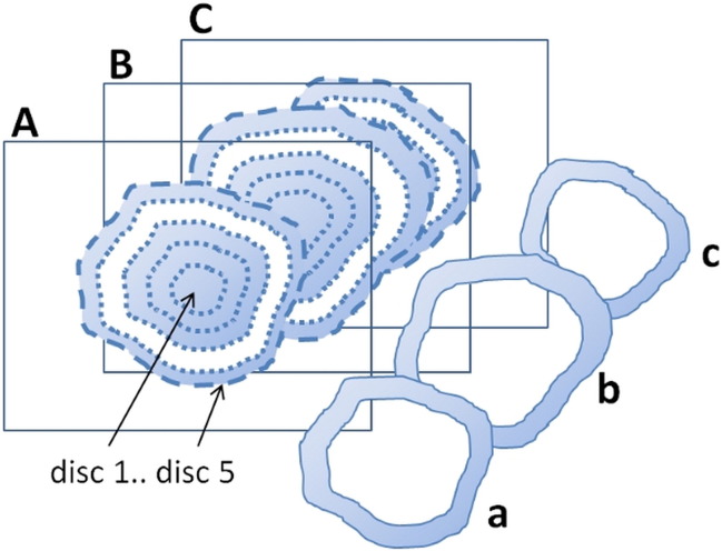
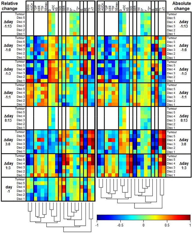
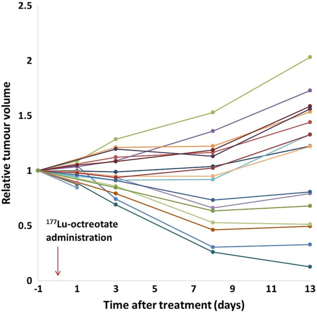
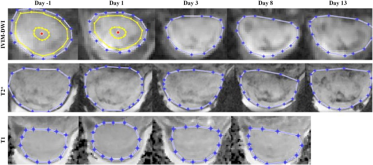
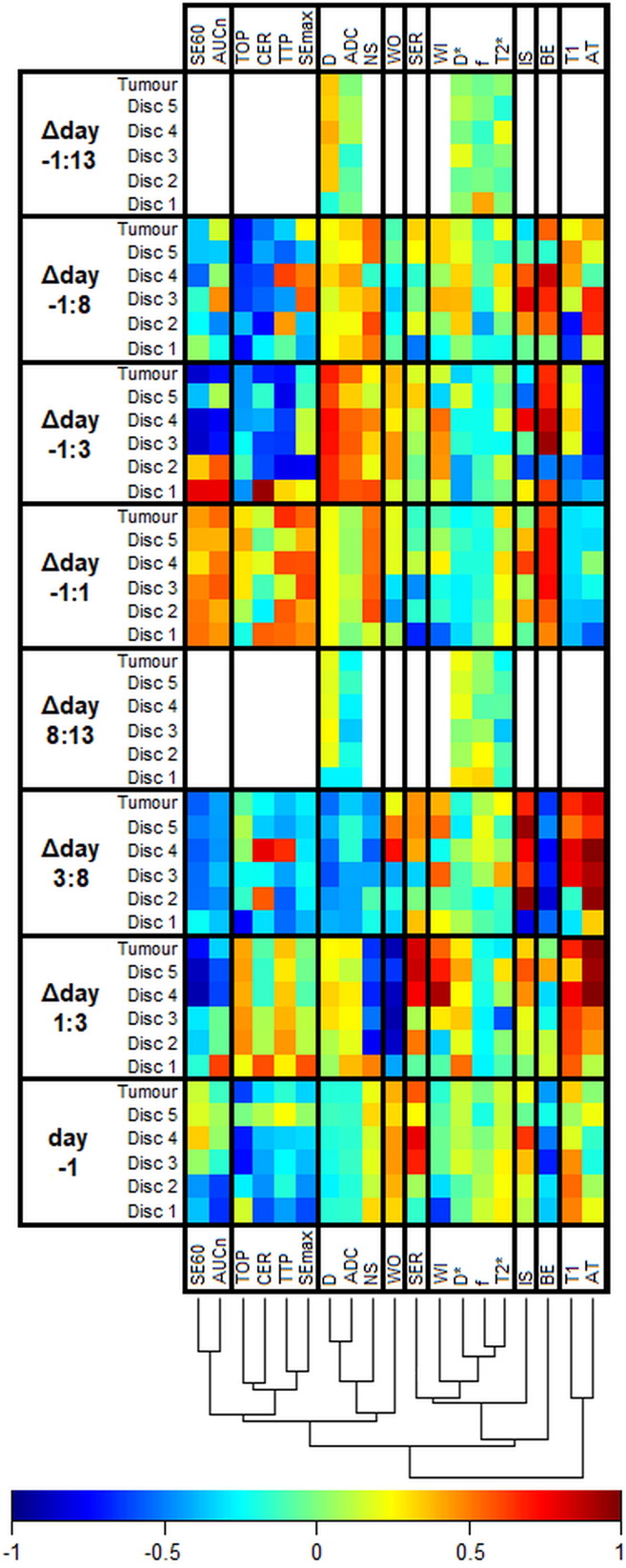
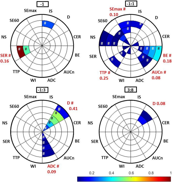
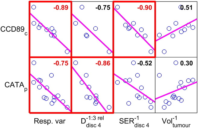
Magnetic resonance (MR) methods enable noninvasive, regional tumor therapy response assessment, but associations between MR parameters, underlying biology, and therapeutic effects must be investigated. The aim of this study was to investigate response assessment efficacy and biological associations of MR parameters in a neuroendocrine tumor (NET) model subjected to radionuclide treatment. Twenty-one mice with NETs received Lu-octreotate at day 0. MR experiments (day -1, 1, 3, 8, and 13) included T2-weighted, dynamic contrast-enhanced (DCE) and diffusion-weighted imaging (DWI) and relaxation measurements (T1/T2*). Tumor tissue was analyzed using proteomics. MR-derived parameters were evaluated for each examination day and for different radial distances from the tumor center. Response assessment efficacy and biological associations were evaluated using feature selection and protein expression correlations, respectively. Reduced tumor growth rate or shrinkage was observed until day 8, followed by reestablished growth in most tumors. The most important MR parameter for response prediction was DCE-MRI-derived pretreatment signal enhancement ratio (SER) at 40% to 60% radial distance, where it correlated significantly also with centrally sampled protein CCD89 (association: DNA damage and repair, proliferation, cell cycle arrest). The second most important was changed diffusion (D) between day -1 and day 3, at 60% to 80% radial distance, where it correlated significantly also with peripherally sampled protein CATA (association: oxidative stress, proliferation, cell cycle arrest, apoptotic cell death). Important information regarding tumor biology in response to radionuclide therapy is reflected in several MR parameters, SER and D in particular. The spatial and temporal information provided by MR methods increases the sensitivity for tumor therapy response.
磁共振(MR)方法能够实现无创的区域肿瘤治疗反应评估,但必须研究MR参数、潜在生物学特性与治疗效果之间的关联。本研究的目的是在接受放射性核素治疗的神经内分泌肿瘤(NET)模型中,研究MR参数的反应评估效能及生物学关联。21只患有NET的小鼠在第0天接受了镥-奥曲肽治疗。MR实验(第-1天、第1天、第3天、第8天和第13天)包括T2加权成像、动态对比增强(DCE)成像和扩散加权成像(DWI)以及弛豫测量(T1/T2*)。使用蛋白质组学分析肿瘤组织。对每个检查日以及距肿瘤中心不同径向距离处的MR衍生参数进行评估。分别使用特征选择和蛋白质表达相关性评估反应评估效能和生物学关联。在第8天之前观察到肿瘤生长速率降低或缩小,随后大多数肿瘤重新开始生长。用于反应预测的最重要MR参数是在径向距离40%至60%处DCE-MRI衍生的治疗前信号增强率(SER),在此处它还与中央采样的蛋白质CCD89显著相关(关联:DNA损伤与修复、增殖、细胞周期停滞)。第二重要的是在第-1天和第3天之间、径向距离60%至80%处的扩散(D)变化,在此处它还与外周采样的蛋白质CATA显著相关(关联:氧化应激、增殖、细胞周期停滞、凋亡性细胞死亡)。关于放射性核素治疗反应中肿瘤生物学的重要信息反映在几个MR参数中,尤其是SER和D。MR方法提供的空间和时间信息提高了肿瘤治疗反应的敏感性。