Institute of Clinical Sciences, Sahlgrenska Cancer Center, Sahlgrenska Academy, Department of Radiation Physics, University of Gothenburg, Gothenburg, Sweden.
Department of Medical Physics and Biomedical Engineering, Sahlgrenska University Hospital, Gothenburg, Sweden.
NMR Biomed. 2019 Mar;32(3):e4060. doi: 10.1002/nbm.4060. Epub 2019 Jan 28.
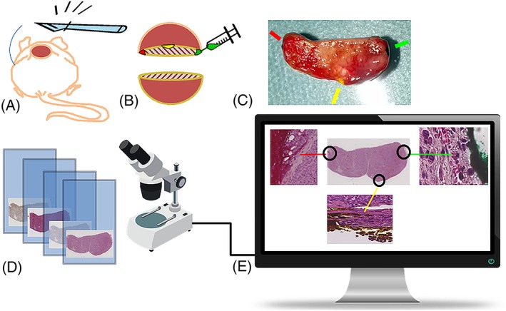
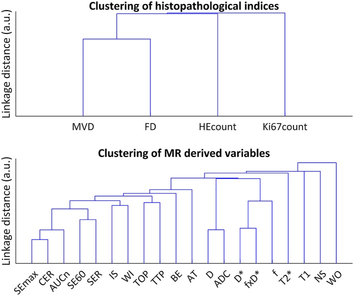
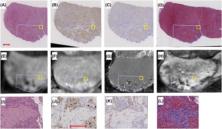
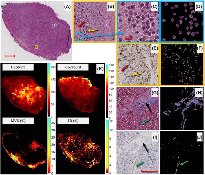
Early non-invasive tumour therapy response assessment requires methods sensitive to biological and physiological tumour characteristics. The aim of this study was to find and evaluate magnetic resonance imaging (MRI) derived tumour tissue parameters that correlate with histological parameters and that reflect effects of radionuclide therapy. Mice bearing a subcutaneous human small-intestine neuroendocrine tumour were i.v. injected with Lu-octreotate. MRI was performed (7 T Bruker Biospec) on different post-therapy intervals (1 and 13 days) using T2-weighted imaging, mapping of T2* and T1 relaxation time constants, as well as diffusion and dynamic contrast enhancement (DCE-MRI) techniques. After MRI, animals were killed and tumours excised. Four differently stained histological sections of the most central imaged tumour plane were digitized, and segmentation techniques were used to produce maps reflecting fibrotic and vascular density, apoptosis, and proliferation. Histological maps were aligned with MRI-derived parametric maps using landmark-based registration. Correlations and predictive power were evaluated using linear mixed-effects models and cross-validation, respectively. Several MR parameters showed statistically significant correlations with histological parameters. In particular, three DCE-MRI-derived parameters reflecting capillary function additionally showed high predictive power regarding apoptosis (2/3) and proliferation (1/3). T1 could be used to predict vascular density, and perfusion fraction derived from diffusion MRI could predict fibrotic density, although with lower predictive power. This work demonstrates the potential to use multiparametric MRI to retrieve important information on the tumour microenvironment after radiotherapy. The non-invasiveness of the method also allows longitudinal tumour tissue characterization. Further investigation is warranted to evaluate the parameters highlighted in this study longitudinally, in larger studies, and with additional histological methods.
早期无创肿瘤治疗反应评估需要对肿瘤的生物学和生理学特征敏感的方法。本研究旨在寻找和评估与组织学参数相关且能反映放射性核素治疗效果的磁共振成像(MRI)衍生的肿瘤组织参数。皮下植入人小肠神经内分泌肿瘤的小鼠静脉注射 Lu-octreotate。使用 T2 加权成像、T2*和 T1 弛豫时间常数映射以及扩散和动态对比增强(DCE-MRI)技术,在不同的治疗后间隔(1 天和 13 天)对 MRI 进行成像(7T Bruker Biospec)。MRI 后,处死动物并切除肿瘤。对最中心成像肿瘤平面的四个不同染色的组织学切片进行数字化处理,并使用分割技术生成反映纤维化和血管密度、细胞凋亡和增殖的图谱。使用基于标志的配准将组织学图谱与 MRI 衍生的参数图谱对齐。使用线性混合效应模型和交叉验证分别评估相关性和预测能力。几种 MR 参数与组织学参数显示出统计学上的显著相关性。特别是,反映毛细血管功能的三个 DCE-MRI 衍生参数对细胞凋亡(2/3)和增殖(1/3)具有较高的预测能力。T1 可用于预测血管密度,而从扩散 MRI 获得的灌注分数可预测纤维化密度,尽管预测能力较低。这项工作证明了使用多参数 MRI 从放射治疗后获取肿瘤微环境重要信息的潜力。该方法的非侵入性还允许对肿瘤组织进行纵向特征描述。需要进一步研究来评估本研究中突出的参数的纵向、在更大的研究中以及使用其他组织学方法的预测能力。