Department of Neurology, Beijing Hospital, National Center of Gerontology, Beijing 100730, China.
Department of Radiology, Beijing Hospital, National Center of Gerontology, Beijing 100730, China.
Chin Med J (Engl). 2018 Jan 20;131(2):161-170. doi: 10.4103/0366-6999.222322.
Chronic stress contributes to increased risks of atherosclerotic diseases including heart disease, stroke, and transient ischemic attack. However, its underline mechanisms are poorly understood. This study aimed to elucidate the mechanism via which chronic stress exerts its effect on atherosclerosis (AS).
Fifty male New Zealand white rabbits were used. Aortic balloon-injury model was applied. Both social stress and physical stress methods were adopted to establish chronic stress models. The lumen stenotic degree, intimal and medial areas, maximum fibrous cap thickness, and plaque contents were measured with histological sections. Proteomic methods were applied to detect protein changes in abdominal aortas to identify the specialized mediators. Real-time reverse transcription-polymerase chain reaction was used for further verification and investigation.
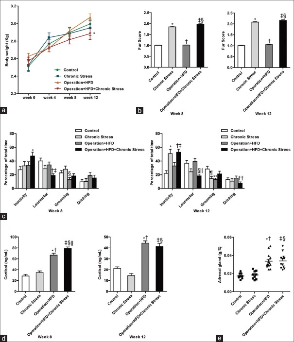
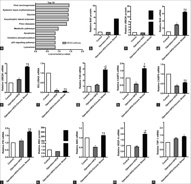
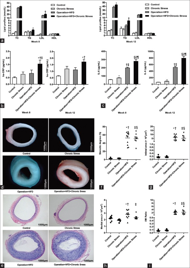
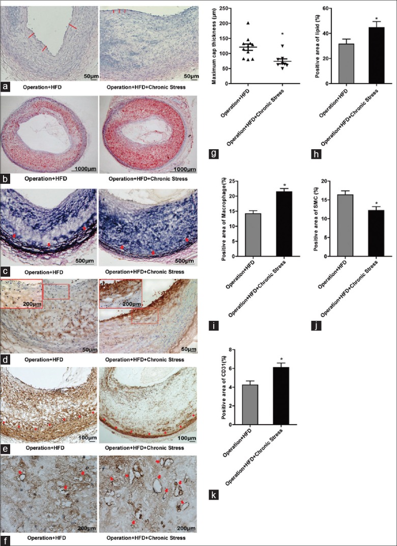
The stress rabbits exhibited lower body weight, worse fur state, more inactivity behavior, and higher serum cortisol level. Chronic stress was significantly associated with the decreased medial area and increased plaque instability, which was manifested by thinner fibrous caps, larger lipid cores, more macrophages, and new vessels but fewer smooth muscle cells and elastic fibers. After chronic stress, the apoptosis-related genes UBE2K, BAX, FAS, Caspase 3, Caspase 9, and P53 were upregulated, and BCL-2/BAX was down-regulated; the angiogenesis-related genes ANG and VEGF-A were also highly expressed in atherosclerotic arteries.
Rabbit models of chronic stress were successfully established by applying both social stress and physical stress for 8 weeks. Chronic stress can reduce AS tunica media and accelerate plaque instability by promoting apoptosis and neovascularization.
慢性应激会增加动脉粥样硬化疾病(包括心脏病、中风和短暂性脑缺血发作)的风险。然而,其潜在机制尚不清楚。本研究旨在阐明慢性应激对动脉粥样硬化(AS)产生影响的机制。
使用 50 只雄性新西兰白兔。应用主动脉球囊损伤模型。采用社会应激和躯体应激方法建立慢性应激模型。通过组织学切片测量管腔狭窄程度、内膜和中膜面积、最大纤维帽厚度和斑块含量。采用蛋白质组学方法检测腹主动脉蛋白变化,以鉴定特异性介质。实时逆转录-聚合酶链反应用于进一步验证和研究。
应激兔体重下降,皮毛状态较差,活动减少,血清皮质醇水平升高。慢性应激与中膜面积减少和斑块不稳定性增加显著相关,表现为纤维帽变薄、脂质核增大、巨噬细胞和新生血管增多、平滑肌细胞和弹性纤维减少。慢性应激后,凋亡相关基因 UBE2K、BAX、FAS、Caspase 3、Caspase 9 和 P53 上调,BCL-2/BAX 下调;血管生成相关基因 ANG 和 VEGF-A 在动脉粥样硬化血管中也高表达。
通过 8 周的社会应激和躯体应激,成功建立了慢性应激兔模型。慢性应激可通过促进细胞凋亡和新生血管形成,减少 AS 中膜并加速斑块不稳定。