Gautam Mukesh, Bhattacharya Indrashis, Rai Umesh, Majumdar Subeer S
Department of Zoology, University of Delhi, Delhi, India.
Cellular Endocrinology Laboratory, National Institute of Immunology, New Delhi, India.
PLoS One. 2018 Jan 17;13(1):e0191201. doi: 10.1371/journal.pone.0191201. eCollection 2018.
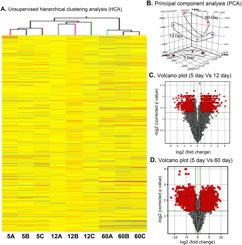
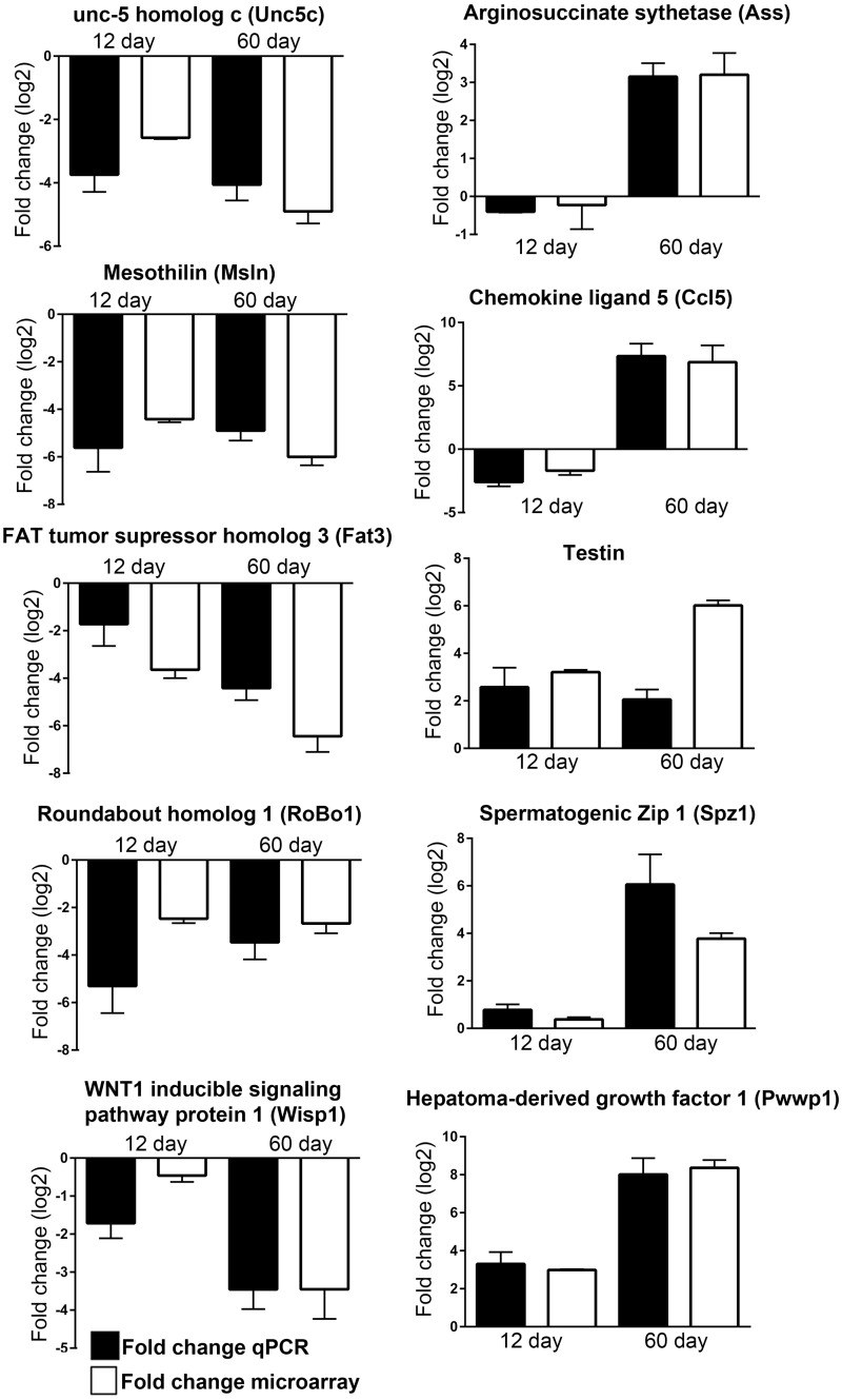
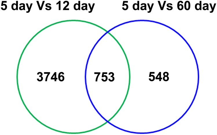
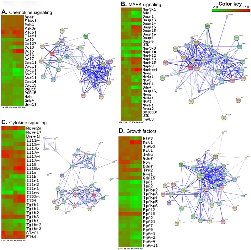
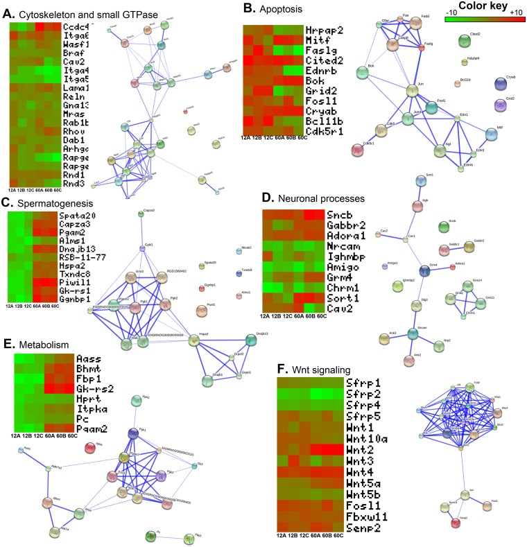
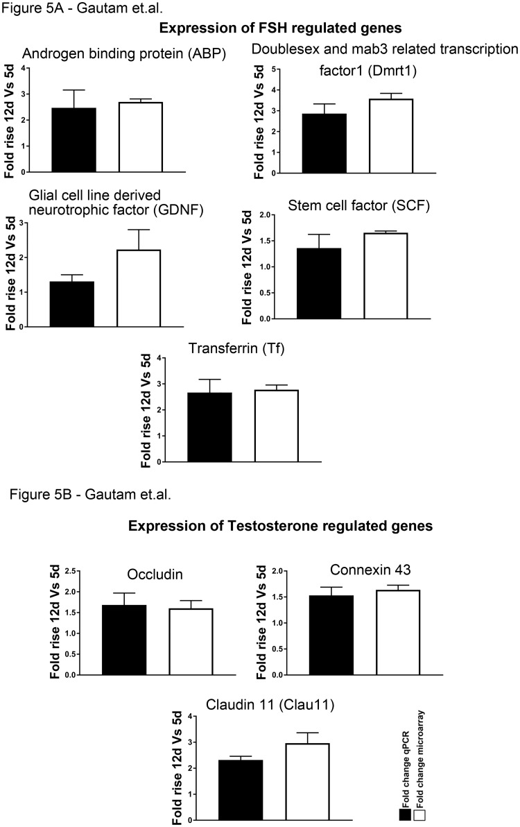
Sertoli cells (Sc) are unique somatic cells of testis that are the target of both FSH and testosterone (T) and regulate spermatogenesis. Although Sc of neonatal rat testes are exposed to high levels of FSH and T, robust differentiation of spermatogonial cells becomes conspicuous only after 11-days of postnatal age. We have demonstrated earlier that a developmental switch in terms of hormonal responsiveness occurs in rat Sc at around 12 days of postnatal age during the rapid transition of spermatogonia A to B. Therefore, such "functional maturation" of Sc, during pubertal development becomes prerequisite for the onset of spermatogenesis. However, a conspicuous difference in robust hormone (both T and FSH) induced gene expression during the different phases of Sc maturation restricts our understanding about molecular events necessary for the spermatogenic onset and maintenance. Here, using microarray technology, we for the first time have compared the differential transcriptional profile of Sc isolated and cultured from immature (5 days old), maturing (12 days old) and mature (60 days old) rat testes. Our data revealed that immature Sc express genes involved in cellular growth, metabolism, chemokines, cell division, MAPK and Wnt pathways, while mature Sc are more specialized expressing genes involved in glucose metabolism, phagocytosis, insulin signaling and cytoskeleton structuring. Taken together, this differential transcriptome data provide an important resource to reveal the molecular network of Sc maturation which is necessary to govern male germ cell differentiation, hence, will improve our current understanding of the etiology of some forms of idiopathic male infertility.
支持细胞(Sc)是睾丸特有的体细胞,是促卵泡激素(FSH)和睾酮(T)的作用靶点,并调节精子发生。尽管新生大鼠睾丸的支持细胞暴露于高水平的FSH和T,但精原细胞的强烈分化仅在出生后11天之后才变得明显。我们之前已经证明,在精原细胞A向B快速转变期间,出生后约12天大鼠支持细胞在激素反应性方面发生了发育转变。因此,支持细胞在青春期发育期间的这种“功能成熟”成为精子发生开始的先决条件。然而,在支持细胞成熟的不同阶段,激素(T和FSH)诱导的基因表达存在明显差异,这限制了我们对精子发生开始和维持所必需的分子事件的理解。在这里,我们首次使用微阵列技术比较了从未成熟(5日龄)、成熟中(12日龄)和成熟(60日龄)大鼠睾丸中分离和培养的支持细胞的差异转录谱。我们的数据显示,未成熟的支持细胞表达参与细胞生长、代谢、趋化因子、细胞分裂、丝裂原活化蛋白激酶(MAPK)和Wnt信号通路的基因,而成熟的支持细胞则更特异地表达参与葡萄糖代谢、吞噬作用、胰岛素信号传导和细胞骨架构建的基因。综上所述,这些差异转录组数据为揭示支持细胞成熟的分子网络提供了重要资源,这对于控制雄性生殖细胞分化是必要的,因此,将改善我们目前对某些形式的特发性男性不育病因的理解。