• 文献检索
  • 文档翻译
  • 深度研究
  • 学术资讯
  • Suppr Zotero 插件Zotero 插件
  • 邀请有礼
  • 套餐&价格
  • 历史记录
应用&插件
Suppr Zotero 插件Zotero 插件浏览器插件Mac 客户端Windows 客户端微信小程序
定价
高级版会员购买积分包购买API积分包
服务
文献检索文档翻译深度研究API 文档MCP 服务
关于我们
关于 Suppr公司介绍联系我们用户协议隐私条款
关注我们

Suppr 超能文献

核心技术专利:CN118964589B侵权必究
粤ICP备2023148730 号-1Suppr @ 2026

文献检索

告别复杂PubMed语法,用中文像聊天一样搜索,搜遍4000万医学文献。AI智能推荐,让科研检索更轻松。

立即免费搜索

文件翻译

保留排版,准确专业,支持PDF/Word/PPT等文件格式,支持 12+语言互译。

免费翻译文档

深度研究

AI帮你快速写综述,25分钟生成高质量综述,智能提取关键信息,辅助科研写作。

立即免费体验

S1 骶髂螺钉进针点与臀上动脉之间的解剖关系。

Anatomical relation between S1 sacroiliac screws' entrance points and superior gluteal artery.

作者信息

Zhao Yong, You Libo, Lian Wei, Zou Dexin, Dong Shengjie, Sun Tao, Zhang Shudong, Wang Dan, Li Jingning, Li Wenliang, Zhao Yuchi

机构信息

Orthopaedics Department, Yantai Shan Hospital, 91#, Jiefang Road, Yantai, 264008, Shandong Province, People's Republic of China.

Operating Room, Yantai Shan Hospital, 91#, Jiefang Road, Yantai, 264008, Shandong Province, People's Republic of China.

出版信息

J Orthop Surg Res. 2018 Jan 18;13(1):15. doi: 10.1186/s13018-018-0713-5.

DOI:10.1186/s13018-018-0713-5
PMID:29347992
原文链接:https://pmc.ncbi.nlm.nih.gov/articles/PMC5774034/
Abstract

BACKGROUND

To conduct radiologic anatomical study on the relation between S1 sacroiliac screws' entry points and the route of the pelvic outer superior gluteal artery branches with the aim to provide the anatomical basis and technical reference for the avoidance of damage to the superior gluteal artery during the horizontal sacroiliac screw placement.

METHODS

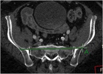
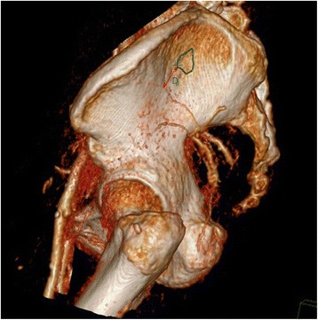
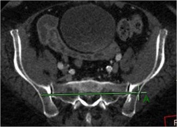
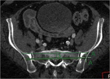
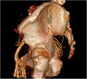
Superior gluteal artery CTA (CT angiography) vascular imaging of 74 healthy adults (37 women and 37 men) was done with 128-slice spiral CT (computed tomography). The CT attendant-measuring software was used to portray the "safe bony entrance area" (hereinafter referred to as "Safe Area") of the S1 segment in the standard lateral pelvic view of three-dimensional reconstruction. The anatomical relation between S1 sacroiliac screws' Safe Area and the pelvic outer superior gluteal artery branches was observed and recorded. The number of cases in which artery branches intersected the Safe Area was counted. The cases in which superior gluteal artery branches disjointed from the Safe Area were identified, and the shortest distance between the Safe Area and the superior gluteal artery branch closest to the Safe Area was measured.

RESULTS

Three cases out of the 74 sample cases were excluded from this study as they were found to have no bony space for horizontal screw placement in S1 segment. Among the remaining 71 sample cases, there are 32 cases (45.1%) where the deep superior branch of superior gluteal artery passes through the Safe Area of S1 entrance point. There was no distinguishing feature and rule on how the deep superior branches and the Safe Area overlapped. In the 39 cases in which superior gluteal artery branches disjointed from the Safe Area, the deep superior branches of superior gluteal artery were the branches closest to the Safe Area and the part of the branch closest to the Safe Area was located in front of the widest part of the Safe Area. The shortest distance between the deep superior branch and the Safe Area is 0.86 ± 0.84 cm.

CONCLUSION

There is a high risk of accidental injury of the deep superior branches of superior gluteal artery in the process of S1 sacroiliac screw placement. Even if the entry points are located in the safe bony entrance area, the absolute secure placement cannot be assured. We suggest that great attention should be paid to make thorough preoperative plans.

摘要

背景

对S1骶髂螺钉进针点与臀上动脉外侧支走行的关系进行影像学解剖学研究,旨在为骶髂螺钉水平置入时避免损伤臀上动脉提供解剖学依据和技术参考。

方法

采用128层螺旋CT对74例健康成年人(37例女性,37例男性)进行臀上动脉CT血管造影(CTA)血管成像。利用CT随诊测量软件在三维重建的骨盆标准侧位视图中描绘S1节段的“安全骨性入路区域”(以下简称“安全区”)。观察并记录S1骶髂螺钉安全区与臀上动脉外侧支的解剖关系。统计动脉分支与安全区相交的病例数。识别臀上动脉分支与安全区不相交的病例,并测量安全区与最靠近安全区的臀上动脉分支之间的最短距离。

结果

74例样本病例中有3例因S1节段无水平螺钉置入的骨性空间而被排除在本研究之外。在其余71例样本病例中,有32例(45.1%)臀上动脉深上支穿过S1进针点的安全区。深上支与安全区重叠的方式无明显特征和规律。在39例臀上动脉分支与安全区不相交的病例中,臀上动脉深上支是最靠近安全区的分支,且分支最靠近安全区的部分位于安全区最宽部分的前方。深上支与安全区之间的最短距离为0.86±0.84cm。

结论

S1骶髂螺钉置入过程中臀上动脉深上支意外损伤风险较高。即使进针点位于安全骨性入路区域,也不能保证绝对安全置入。建议术前应高度重视并制定详尽的计划。

https://cdn.ncbi.nlm.nih.gov/pmc/blobs/8b59/5774034/4c5245f8ed79/13018_2018_713_Fig5_HTML.jpg
https://cdn.ncbi.nlm.nih.gov/pmc/blobs/8b59/5774034/916904c9a5df/13018_2018_713_Fig1_HTML.jpg
https://cdn.ncbi.nlm.nih.gov/pmc/blobs/8b59/5774034/c123ed70501f/13018_2018_713_Fig2_HTML.jpg
https://cdn.ncbi.nlm.nih.gov/pmc/blobs/8b59/5774034/db0e7da498af/13018_2018_713_Fig3_HTML.jpg
https://cdn.ncbi.nlm.nih.gov/pmc/blobs/8b59/5774034/8edb46adfbad/13018_2018_713_Fig4_HTML.jpg
https://cdn.ncbi.nlm.nih.gov/pmc/blobs/8b59/5774034/4c5245f8ed79/13018_2018_713_Fig5_HTML.jpg

相似文献

1
Anatomical relation between S1 sacroiliac screws' entrance points and superior gluteal artery.S1 骶髂螺钉进针点与臀上动脉之间的解剖关系。
J Orthop Surg Res. 2018 Jan 18;13(1):15. doi: 10.1186/s13018-018-0713-5.
2
Anatomic relationship between S2 sacroiliac screws' entry points and pelvic external branches of superior gluteal artery.S2 骶髂螺钉进钉点与臀上动脉骨盆外支的解剖关系。
Eur J Trauma Emerg Surg. 2022 Apr;48(2):857-862. doi: 10.1007/s00068-021-01622-2. Epub 2021 Mar 6.
3
Parameters of lengthened sacroiliac screw fixation: a radiological anatomy study.延长骶髂螺钉固定的参数:一项影像学解剖研究。
Eur Spine J. 2012 Sep;21(9):1807-14. doi: 10.1007/s00586-012-2367-z. Epub 2012 May 18.
4
Superior gluteal artery injury during iliosacral screw placement due to aberrant anatomy.由于解剖结构异常,在置入髂骶螺钉过程中臀上动脉损伤。
Orthopedics. 2010 Feb;33(2):117-20. doi: 10.3928/01477447-20100104-26.
5
Risks to the superior gluteal neurovascular bundle during percutaneous iliosacral screw insertion: an anatomical cadaver study.经皮髂骶螺钉置入过程中臀上神经血管束的风险:一项解剖学尸体研究
J Orthop Trauma. 2005 Feb;19(2):96-101. doi: 10.1097/00005131-200502000-00005.
6
Evaluation of Partial Cut-out of Sacroiliac Screws From the Sacral Ala Slope via Pelvic Inlet and Outlet View.通过骨盆入口和出口位评估骶髂螺钉从骶骨翼斜坡部分穿出的情况。
Spine (Phila Pa 1976). 2015 Dec;40(24):E1264-8. doi: 10.1097/BRS.0000000000001134.
7
Risks to the Superior Gluteal Neurovascular Bundle During Iliosacral and Transsacral Screw Fixation: A Computed Tomogram Arteriography Study.髂骶和经骶骨螺钉固定期间臀上神经血管束的风险:一项计算机断层血管造影研究
J Orthop Trauma. 2017 Dec;31(12):640-643. doi: 10.1097/BOT.0000000000000996.
8
The influence of sacral morphology on the existence of secure S1 and S2 transverse bone corridors for iliosacroiliac screw fixation.骶骨形态对用于髂骶螺钉固定的安全S1和S2横向骨通道存在情况的影响。
Injury. 2013 Dec;44(12):1773-9. doi: 10.1016/j.injury.2013.08.006. Epub 2013 Aug 14.
9
[A morphologic evaluation of the sacroiliac joint and plate fixation on a pelvic model using a S1 pedicular screw, transiliosacral screws, and a compression rod for sacroiliac joint injuries].[使用S1椎弓根螺钉、经髂骶螺钉和加压棒对骨盆模型上的骶髂关节及钢板固定进行形态学评估以治疗骶髂关节损伤]
Acta Orthop Traumatol Turc. 2002;36(5):432-41.
10
Topographic localization of the sacroiliac joint and superior gluteal artery branches on the posterolateral ilium.髂后上棘的骶髂关节和臀上动脉分支的定位。
Clin Anat. 2023 Oct;36(7):971-976. doi: 10.1002/ca.24005. Epub 2023 Jan 24.

引用本文的文献

1
CT Angiographic Evaluation of Superior Gluteal Artery Branches in Lateral Sacroiliac Joint Fusion: A Pilot Study to Inform Preoperative Surgical Planning and Anatomic Parameters.骶髂关节外侧融合术中臀上动脉分支的CT血管造影评估:一项为术前手术规划和解剖参数提供信息的初步研究
J Pain Res. 2025 Jun 19;18:3069-3075. doi: 10.2147/JPR.S514778. eCollection 2025.
2
Preoperative 3D printing planning technology combined with orthopedic surgical robot-assisted minimally invasive screw fixation for the treatment of pelvic fractures: a retrospective study.术前三维打印规划技术联合骨科手术机器人辅助微创螺钉内固定治疗骨盆骨折:一项回顾性研究
PeerJ. 2024 Dec 12;12:e18632. doi: 10.7717/peerj.18632. eCollection 2024.
3

本文引用的文献

1
Comparison of the risk of breakage of two kinds of sacroiliac screws in the treatment of bilateral sacral fractures.两种骶髂螺钉治疗双侧骶骨骨折时的断裂风险比较
Eur Spine J. 2014 Jul;23(7):1558-67. doi: 10.1007/s00586-014-3313-z. Epub 2014 Apr 27.
2
Mechanical comparison between lengthened and short sacroiliac screws in sacral fracture fixation: a finite element analysis.骶骨骨折固定中加长与短型骶髂螺钉的力学比较:有限元分析。
Orthop Traumatol Surg Res. 2013 Sep;99(5):601-6. doi: 10.1016/j.otsr.2013.03.023. Epub 2013 Jul 12.
3
Parameters of lengthened sacroiliac screw fixation: a radiological anatomy study.
Safety and Preliminary Effectiveness of Lateral Transiliac Sacroiliac Joint Fusion by Interventional Pain Physicians: A Retrospective Analysis.
介入疼痛科医生经皮侧方骶髂关节融合术的安全性及初步疗效:一项回顾性分析
J Pain Res. 2024 Jun 17;17:2147-2153. doi: 10.2147/JPR.S462072. eCollection 2024.
4
Peroneal artery injury potential due to different syndesmosis screw placement options: a simulation study with lower extremity computed tomography angiography.不同下胫腓联合螺钉固定位置导致腓动脉损伤的潜在风险:下肢 CT 血管造影模拟研究。
Arch Orthop Trauma Surg. 2024 May;144(5):2119-2125. doi: 10.1007/s00402-024-05258-w. Epub 2024 Mar 16.
5
Improving the Stability of a Hemipelvic Prosthesis Based on Bone Mineral Density Screw Channel and Prosthesis Optimization Design.基于骨密度螺钉通道和假体优化设计提高半骨盆假体的稳定性
Front Bioeng Biotechnol. 2022 May 30;10:892385. doi: 10.3389/fbioe.2022.892385. eCollection 2022.
6
Delayed lower extremity paresis following iliosacral screws: Atypical complication and treatment.髂骶螺钉置入术后延迟性下肢轻瘫:一种非典型并发症及治疗方法
Trauma Case Rep. 2020 Dec 10;31:100380. doi: 10.1016/j.tcr.2020.100380. eCollection 2021 Feb.
延长骶髂螺钉固定的参数:一项影像学解剖研究。
Eur Spine J. 2012 Sep;21(9):1807-14. doi: 10.1007/s00586-012-2367-z. Epub 2012 May 18.
4
Comparison of stability of two kinds of sacro-iliac screws in the fixation of bilateral sacral fractures in a finite element model.两种骶骨螺钉在有限元模型中固定双侧骶骨骨折的稳定性比较。
Injury. 2012 Apr;43(4):490-4. doi: 10.1016/j.injury.2011.12.023. Epub 2012 Jan 26.
5
Superior gluteal artery injury during iliosacral screw placement due to aberrant anatomy.由于解剖结构异常,在置入髂骶螺钉过程中臀上动脉损伤。
Orthopedics. 2010 Feb;33(2):117-20. doi: 10.3928/01477447-20100104-26.
6
Pseudoaneurysm of the superior gluteal artery during iliosacral screw fixation.髂骶螺钉固定过程中臀上动脉假性动脉瘤
Acta Orthop Belg. 2007 Aug;73(4):544-7.
7
Risks to the superior gluteal neurovascular bundle during percutaneous iliosacral screw insertion: an anatomical cadaver study.经皮髂骶螺钉置入过程中臀上神经血管束的风险:一项解剖学尸体研究
J Orthop Trauma. 2005 Feb;19(2):96-101. doi: 10.1097/00005131-200502000-00005.
8
Transsacral versus modified pelvic landmarks for percutaneous iliosacral screw placement--a computed tomographic analysis and cadaveric study.经骶骨与改良骨盆标志点用于经皮髂骶螺钉置入的比较——一项CT分析及尸体研究
Am J Orthop (Belle Mead NJ). 2000 Sep;29(9 Suppl):16-21.
9
Safe placement of S1 and S2 iliosacral screws: the "vestibule" concept.S1和S2骶髂螺钉的安全置入:“前庭”概念
J Orthop Trauma. 2000 May;14(4):264-9. doi: 10.1097/00005131-200005000-00007.
10
Superior gluteal artery injury during iliosacral screw placement.髂骶螺钉置入过程中臀上动脉损伤
J Orthop Trauma. 1999 Mar-Apr;13(3):220-7. doi: 10.1097/00005131-199903000-00011.