• 文献检索
  • 文档翻译
  • 深度研究
  • 学术资讯
  • Suppr Zotero 插件Zotero 插件
  • 邀请有礼
  • 套餐&价格
  • 历史记录
应用&插件
Suppr Zotero 插件Zotero 插件浏览器插件Mac 客户端Windows 客户端微信小程序
定价
高级版会员购买积分包购买API积分包
服务
文献检索文档翻译深度研究API 文档MCP 服务
关于我们
关于 Suppr公司介绍联系我们用户协议隐私条款
关注我们

Suppr 超能文献

核心技术专利:CN118964589B侵权必究
粤ICP备2023148730 号-1Suppr @ 2026

文献检索

告别复杂PubMed语法,用中文像聊天一样搜索,搜遍4000万医学文献。AI智能推荐,让科研检索更轻松。

立即免费搜索

文件翻译

保留排版,准确专业,支持PDF/Word/PPT等文件格式,支持 12+语言互译。

免费翻译文档

深度研究

AI帮你快速写综述,25分钟生成高质量综述,智能提取关键信息,辅助科研写作。

立即免费体验

内皮素-1与增殖性糖尿病视网膜病变膜中的纤维化有关。

Endothelin-1 is associated with fibrosis in proliferative diabetic retinopathy membranes.

作者信息

Chang William, Lajko Michelle, Fawzi Amani A

机构信息

Department of Ophthalmology, Northwestern University, Feinberg School of Medicine, Chicago, IL, United States of America.

出版信息

PLoS One. 2018 Jan 19;13(1):e0191285. doi: 10.1371/journal.pone.0191285. eCollection 2018.

DOI:10.1371/journal.pone.0191285
PMID:29351334
原文链接:https://pmc.ncbi.nlm.nih.gov/articles/PMC5774761/
Abstract

PURPOSE

To characterize the relationship between endothelin-1 and fibrosis in epiretinal membranes in proliferative diabetic retinopathy and explore the role of endothelial-mesenchymal transition in these membranes.

METHODS

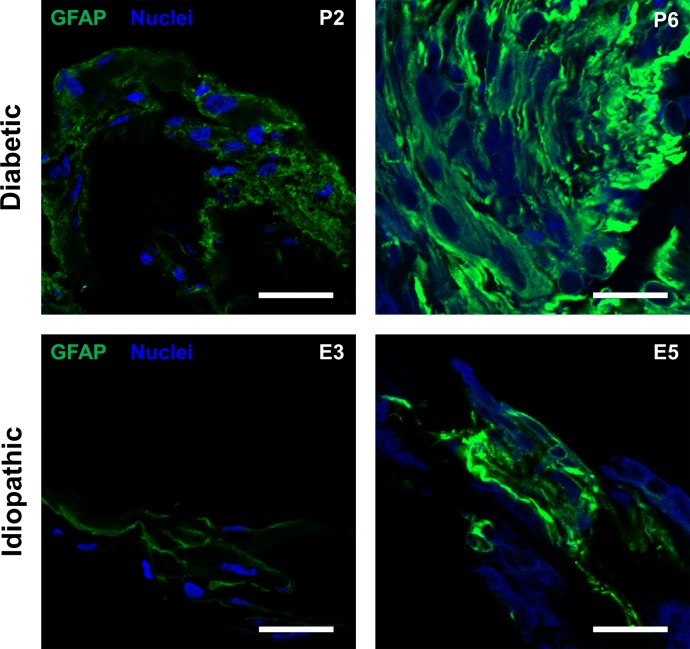
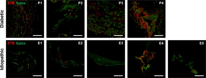
Membranes were obtained from eyes undergoing pars plana vitrectomy for complicated proliferative diabetic retinopathy or idiopathic epiretinal membrane. Through standard immunohistochemical techniques, we labeled membranes to explore the distribution of endothelin-1 and endothelin receptor B, comparing proliferative diabetic retinopathy and idiopathic epiretinal membranes. In addition, membranes were also labeled with markers for fibroblasts, endothelial, and glial cells and studied with confocal laser scanning microscopy. The intensity of endothelin-1 labeling was quantified using standard image analysis software.

RESULTS

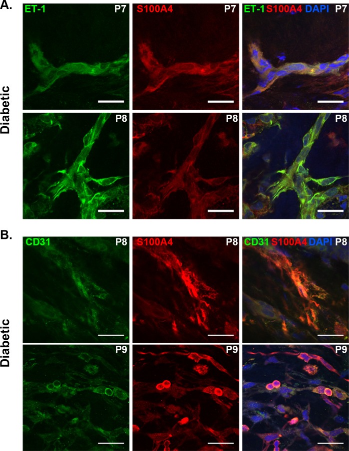
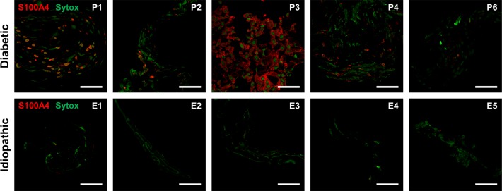
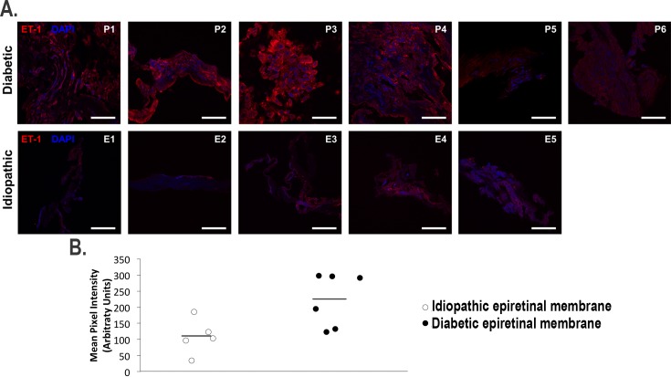
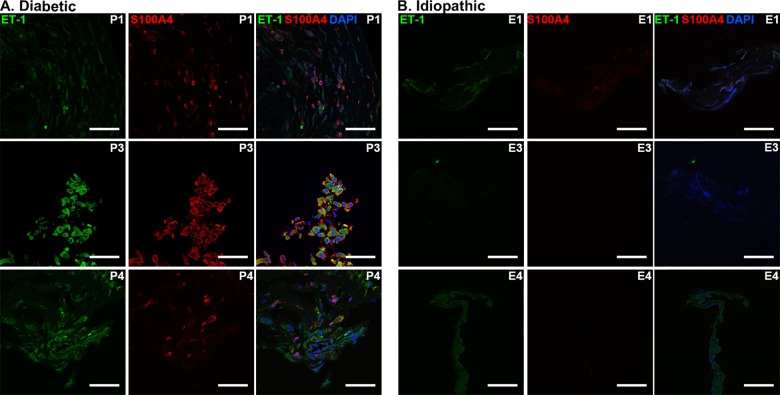
Fourteen membranes were included in the analysis, nine from eyes with proliferative diabetic retinopathy and five idiopathic membranes. Flatmount diabetic membranes showed co-localization of endothelin-1 with S100A4 and CD31. Immunohistochemistry and quantitative analysis of cross-sectional membranes showed significantly higher endothelin-1 labeling in proliferative diabetic retinopathy membranes compared to idiopathic membranes (p<0.05). Diabetic membranes showed more elements staining positive for S100A4 compared to idiopathic membranes.

CONCLUSION

Epiretinal membrane formation in proliferative diabetic retinopathy involves higher tissue levels of endothelin-1 and fibroblastic activity. Furthermore, endothelin-1, endothelial and fibroblastic staining appear to be correlated, suggestive of endothelial-to-mesenchymal transition in proliferative diabetic retinopathy.

摘要

目的

明确增殖性糖尿病视网膜病变视网膜前膜中内皮素-1与纤维化之间的关系,并探讨内皮-间充质转化在这些膜中的作用。

方法

从因复杂性增殖性糖尿病视网膜病变或特发性视网膜前膜而接受玻璃体切割术的眼中获取视网膜前膜。通过标准免疫组织化学技术,标记视网膜前膜以探究内皮素-1和内皮素受体B的分布,比较增殖性糖尿病视网膜病变和特发性视网膜前膜。此外,还用成纤维细胞、内皮细胞和神经胶质细胞的标志物标记视网膜前膜,并采用共聚焦激光扫描显微镜进行研究。使用标准图像分析软件对内皮素-1标记强度进行定量分析。

结果

14个视网膜前膜纳入分析,其中9个来自增殖性糖尿病视网膜病变患者的眼,5个为特发性视网膜前膜。糖尿病视网膜前膜铺片显示内皮素-1与S100A4和CD31共定位。视网膜前膜横断面的免疫组织化学和定量分析显示,增殖性糖尿病视网膜病变视网膜前膜中的内皮素-1标记显著高于特发性视网膜前膜(p<0.05)。与特发性视网膜前膜相比,糖尿病视网膜前膜显示更多S100A4染色阳性成分。

结论

增殖性糖尿病视网膜病变中视网膜前膜的形成涉及更高水平的组织内皮素-1和成纤维细胞活性。此外,内皮素-1、内皮细胞和成纤维细胞染色似乎相关,提示增殖性糖尿病视网膜病变中存在内皮-间充质转化。

https://cdn.ncbi.nlm.nih.gov/pmc/blobs/808b/5774761/182f896e3cdc/pone.0191285.g006.jpg
https://cdn.ncbi.nlm.nih.gov/pmc/blobs/808b/5774761/1b50e9bfc397/pone.0191285.g001.jpg
https://cdn.ncbi.nlm.nih.gov/pmc/blobs/808b/5774761/88a6d3e2b292/pone.0191285.g002.jpg
https://cdn.ncbi.nlm.nih.gov/pmc/blobs/808b/5774761/b2f18260b60b/pone.0191285.g003.jpg
https://cdn.ncbi.nlm.nih.gov/pmc/blobs/808b/5774761/d62777afa05d/pone.0191285.g004.jpg
https://cdn.ncbi.nlm.nih.gov/pmc/blobs/808b/5774761/a84071b8348c/pone.0191285.g005.jpg
https://cdn.ncbi.nlm.nih.gov/pmc/blobs/808b/5774761/182f896e3cdc/pone.0191285.g006.jpg

相似文献

1
Endothelin-1 is associated with fibrosis in proliferative diabetic retinopathy membranes.内皮素-1与增殖性糖尿病视网膜病变膜中的纤维化有关。
PLoS One. 2018 Jan 19;13(1):e0191285. doi: 10.1371/journal.pone.0191285. eCollection 2018.
2
A comparison of hypoxia-inducible factor-α in surgically excised neovascular membranes of patients with diabetes compared with idiopathic epiretinal membranes in nondiabetic patients.比较糖尿病患者手术切除的新生血管膜与非糖尿病患者特发性视网膜前膜中的缺氧诱导因子-α。
Retina. 2010 Oct;30(9):1472-8. doi: 10.1097/IAE.0b013e3181d6df09.
3
APOPTOSIS AND ANGIOFIBROSIS IN DIABETIC TRACTIONAL MEMBRANES AFTER VASCULAR ENDOTHELIAL GROWTH FACTOR INHIBITION: Results of a Prospective Trial. Report No. 2.血管内皮生长因子抑制后糖尿病牵引性膜中的细胞凋亡和血管纤维瘤形成:一项前瞻性试验的结果。报告 2 号。
Retina. 2019 Feb;39(2):265-273. doi: 10.1097/IAE.0000000000001952.
4
Expression of wild-type p53-induced phosphatase 1 in diabetic epiretinal membranes.野生型p53诱导的磷酸酶1在糖尿病性视网膜前膜中的表达。
Oncotarget. 2017 May 30;8(22):35532-35541. doi: 10.18632/oncotarget.16683.
5
Myofibroblasts in proliferative diabetic retinopathy can originate from infiltrating fibrocytes and through endothelial-to-mesenchymal transition (EndoMT).增殖性糖尿病视网膜病变中的肌成纤维细胞可起源于浸润的纤维细胞,并通过内皮-间充质转化(EndoMT)产生。
Exp Eye Res. 2015 Mar;132:179-89. doi: 10.1016/j.exer.2015.01.023. Epub 2015 Jan 28.
6
Alphab-crystallin expression in epiretinal membrane of human proliferative diabetic retinopathy.αB-晶状体蛋白在人增生性糖尿病视网膜病变的视网膜前膜中的表达。
Retina. 2012 Jun;32(6):1190-6. doi: 10.1097/IAE.0b013e318233ab9c.
7
Increased expression of placenta growth factor in proliferative diabetic retinopathy.胎盘生长因子在增殖性糖尿病视网膜病变中的表达增加。
Lab Invest. 1998 Jan;78(1):109-16.
8
Immunohistochemical study of extracellular matrix components in epiretinal membranes of vitreoproliferative retinopathy and proliferative diabetic retinopathy.玻璃体增殖性视网膜病变和增殖性糖尿病视网膜病变视网膜前膜细胞外基质成分的免疫组织化学研究
Eur J Ophthalmol. 2005 May-Jun;15(3):384-91. doi: 10.1177/112067210501500312.
9
Recognition of vitreoschisis in proliferative diabetic retinopathy. A useful landmark in vitrectomy for diabetic traction retinal detachment.增殖性糖尿病视网膜病变中玻璃体劈裂的识别。糖尿病性牵引性视网膜脱离玻璃体切除术中一个有用的标志。
Ophthalmology. 1996 Feb;103(2):323-8. doi: 10.1016/s0161-6420(96)30697-0.
10
Vitreous and plasma concentrations of apelin and vascular endothelial growth factor after intravitreal bevacizumab in eyes with proliferative diabetic retinopathy.玻璃体内和血浆中血管紧张素原肽和血管内皮生长因子在增生性糖尿病视网膜病变眼内注射bevacizumab 后的浓度。
Retina. 2011 Jan;31(1):161-8. doi: 10.1097/IAE.0b013e3181e46ad8.

引用本文的文献

1
Insights into the molecular underpinning of type 2 diabetes complications.对2型糖尿病并发症分子基础的见解。
Hum Mol Genet. 2025 Mar 7;34(6):469-480. doi: 10.1093/hmg/ddae203.
2
GLCCI1 alleviates GRP78-initiated endoplasmic reticulum stress-induced apoptosis of retinal ganglion cells in diabetic retinopathy by upregulating and interacting with HSP90AB1.葡萄糖调节蛋白 78 诱导的内质网应激诱导糖尿病视网膜病变中视网膜神经节细胞凋亡,通过上调和相互作用热休克蛋白 90AB1 减轻。
Sci Rep. 2024 Nov 4;14(1):26665. doi: 10.1038/s41598-024-75874-4.
3
Mechanisms of Xuefu Zhuyu Tang in the Treatment of Diabetic Erectile Dysfunction in Rats Through the Regulation of Vascular Endothelial Function by CaSR/PLC/PKC and MEK/ERK/RSK Pathways.

本文引用的文献

1
Fibrocytes and Fibrovascular Membrane Formation in Proliferative Diabetic Retinopathy.增殖性糖尿病视网膜病变中的纤维细胞与纤维血管膜形成
Invest Ophthalmol Vis Sci. 2016 Sep 1;57(11):4999-5005. doi: 10.1167/iovs.16-19798.
2
Endothelial to Mesenchymal Transition (EndoMT) in the Pathogenesis of Human Fibrotic Diseases.内皮-间充质转化(EndoMT)在人类纤维化疾病发病机制中的作用
J Clin Med. 2016 Apr 11;5(4):45. doi: 10.3390/jcm5040045.
3
In Brief: Endothelial-to-mesenchymal transition.简而言之:内皮-间充质转化。
血府逐瘀汤通过 CaSR/PLC/PKC 和 MEK/ERK/RSK 通路调节血管内皮功能治疗糖尿病性勃起功能障碍的机制。
Am J Mens Health. 2024 Sep-Oct;18(5):15579883241277423. doi: 10.1177/15579883241277423.
4
Epiretinal membranes in patients with uveitis: an update on the current state of management.葡萄膜炎患者的视网膜前膜:当前管理现状的更新。
Int Ophthalmol. 2024 Jun 28;44(1):291. doi: 10.1007/s10792-024-03199-2.
5
Transcriptional Comparison of Human and Murine Retinal Neovascularization.人类和鼠类视网膜新生血管转录组比较。
Invest Ophthalmol Vis Sci. 2023 Dec 1;64(15):46. doi: 10.1167/iovs.64.15.46.
6
Choroidal Vascularity Index Fluctuations in Epiretinal Membranes in Vitreoretinal Surgery: Comparison Between Idiopathic and Diabetic Ones.视网膜玻璃体手术中特发性和糖尿病性视网膜前膜的脉络膜血管密度波动比较。
Transl Vis Sci Technol. 2023 Dec 1;12(12):9. doi: 10.1167/tvst.12.12.9.
7
Advances and Perspectives in Relation to the Molecular Basis of Diabetic Retinopathy-A Review.糖尿病视网膜病变分子基础的研究进展与展望——综述
Biomedicines. 2023 Nov 1;11(11):2951. doi: 10.3390/biomedicines11112951.
8
Endothelial-derived extracellular vesicles associated with electronic cigarette use impair cerebral microvascular cell function.与使用电子烟相关的内皮细胞衍生的细胞外囊泡会损害脑血管细胞功能。
J Appl Physiol (1985). 2023 Aug 1;135(2):271-278. doi: 10.1152/japplphysiol.00243.2023. Epub 2023 Jun 22.
9
Diabetic retinopathy as the leading cause of blindness and early predictor of cascading complications-risks and mitigation.糖尿病视网膜病变是失明的主要原因以及一系列并发症的早期预测指标——风险与缓解措施。
EPMA J. 2023 Feb 13;14(1):21-42. doi: 10.1007/s13167-023-00314-8. eCollection 2023 Mar.
10
The integrated transcriptome bioinformatics analysis identifies key genes and cellular components for proliferative diabetic retinopathy.整合转录组生物信息学分析确定增殖性糖尿病视网膜病变的关键基因和细胞成分。
PLoS One. 2022 Nov 21;17(11):e0277952. doi: 10.1371/journal.pone.0277952. eCollection 2022.
J Pathol. 2016 Feb;238(3):378-80. doi: 10.1002/path.4653. Epub 2016 Jan 4.
4
The Endothelial-mesenchymal Transition in Systemic Sclerosis Is Induced by Endothelin-1 and Transforming Growth Factor-β and May Be Blocked by Macitentan, a Dual Endothelin-1 Receptor Antagonist.系统性硬化症中的内皮-间充质转化由内皮素-1和转化生长因子-β诱导,且可能被双重内皮素-1受体拮抗剂马西替坦阻断。
J Rheumatol. 2015 Oct;42(10):1808-16. doi: 10.3899/jrheum.150088. Epub 2015 Aug 15.
5
Characterization of cells from patient-derived fibrovascular membranes in proliferative diabetic retinopathy.增殖性糖尿病视网膜病变患者来源的纤维血管膜细胞的特征分析
Mol Vis. 2015 Jun 12;21:673-87. eCollection 2015.
6
Fibrosis--a common pathway to organ injury and failure.纤维化——器官损伤和衰竭的常见途径。
N Engl J Med. 2015 Mar 19;372(12):1138-49. doi: 10.1056/NEJMra1300575.
7
Myofibroblasts in proliferative diabetic retinopathy can originate from infiltrating fibrocytes and through endothelial-to-mesenchymal transition (EndoMT).增殖性糖尿病视网膜病变中的肌成纤维细胞可起源于浸润的纤维细胞,并通过内皮-间充质转化(EndoMT)产生。
Exp Eye Res. 2015 Mar;132:179-89. doi: 10.1016/j.exer.2015.01.023. Epub 2015 Jan 28.
8
Endothelial-to-mesenchymal transition in pulmonary hypertension.肺高血压中的内皮细胞-间充质转化。
Circulation. 2015 Mar 17;131(11):1006-18. doi: 10.1161/CIRCULATIONAHA.114.008750. Epub 2015 Jan 15.
9
Endothelin-1 upregulation mediates aging-related cardiac fibrosis.内皮素-1 的上调介导与衰老相关的心脏纤维化。
J Mol Cell Cardiol. 2015 Mar;80:101-9. doi: 10.1016/j.yjmcc.2015.01.001. Epub 2015 Jan 10.
10
Mechanisms of endothelial to mesenchymal transition in the retina in diabetes.糖尿病视网膜中内皮向间充质转化的机制
Invest Ophthalmol Vis Sci. 2014 Oct 21;55(11):7321-31. doi: 10.1167/iovs.14-15167.