• 文献检索
  • 文档翻译
  • 深度研究
  • 学术资讯
  • Suppr Zotero 插件Zotero 插件
  • 邀请有礼
  • 套餐&价格
  • 历史记录
应用&插件
Suppr Zotero 插件Zotero 插件浏览器插件Mac 客户端Windows 客户端微信小程序
定价
高级版会员购买积分包购买API积分包
服务
文献检索文档翻译深度研究API 文档MCP 服务
关于我们
关于 Suppr公司介绍联系我们用户协议隐私条款
关注我们

Suppr 超能文献

核心技术专利:CN118964589B侵权必究
粤ICP备2023148730 号-1Suppr @ 2026

文献检索

告别复杂PubMed语法,用中文像聊天一样搜索,搜遍4000万医学文献。AI智能推荐,让科研检索更轻松。

立即免费搜索

文件翻译

保留排版,准确专业,支持PDF/Word/PPT等文件格式,支持 12+语言互译。

免费翻译文档

深度研究

AI帮你快速写综述,25分钟生成高质量综述,智能提取关键信息,辅助科研写作。

立即免费体验

青光眼的视网膜血管系统:综述

Retinal vasculature in glaucoma: a review.

作者信息

Chan Karen K W, Tang Fangyao, Tham Clement C Y, Young Alvin L, Cheung Carol Y

机构信息

Department of Ophthalmology and Visual Sciences, The Chinese University of Hong Kong, Hong Kong, China.

Department of Ophthalmology and Visual Sciences, Prince of Wales Hospital and Alice Ho Miu Ling Nethersole Hospital, Hong Kong, China.

出版信息

BMJ Open Ophthalmol. 2017 Jul 11;1(1):e000032. doi: 10.1136/bmjophth-2016-000032. eCollection 2017.

DOI:10.1136/bmjophth-2016-000032
PMID:29354699
原文链接:https://pmc.ncbi.nlm.nih.gov/articles/PMC5721639/
Abstract

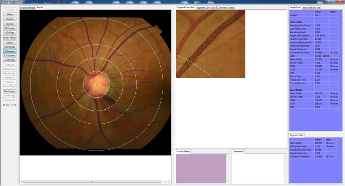
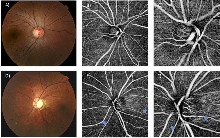
Despite the critical impact of glaucoma on global blindness, its aetiology is not fully characterised. Elevated intraocular pressure is highly associated with glaucomatous optic neuropathy. However, visual field loss still progresses in some patients with normal or even low intraocular pressure. Vascular factors have been suggested to play a role in glaucoma development, based on numerous studies showing associations of glaucoma with blood pressure, ocular perfusion pressure, vasospasm, cardiovascular disease and ocular blood flow. As the retinal vasculature is the only part of the human circulation that readily allows non-invasive visualisation of the microcirculation, a number of quantitative retinal vascular parameters measured from retinal photographs using computer software (eg, calibre, fractal dimension, tortuosity and branching angle) are currently being explored for any association with glaucoma and its progression. Several population-based and clinical studies have reported that changes in retinal vasculature (eg, retinal arteriolar narrowing and decreased fractal dimension) are associated with optic nerve damage and glaucoma, supporting the vascular theory of glaucoma pathogenesis. This review summarises recent findings on the relationships between quantitatively measured structural retinal vascular changes with glaucoma and other markers of optic nerve head damage, including retinal nerve fibre layer thickness. Clinical implications, recent new advances in retinal vascular imaging (eg, optical coherence tomography angiography) and future research directions are also discussed.

摘要

尽管青光眼对全球失明情况有着严重影响,但其病因尚未完全明确。眼压升高与青光眼性视神经病变高度相关。然而,在一些眼压正常甚至偏低的患者中,视野缺损仍会进展。基于大量显示青光眼与血压、眼灌注压、血管痉挛、心血管疾病及眼血流量之间存在关联的研究,血管因素被认为在青光眼的发展过程中起作用。由于视网膜血管系统是人体循环中唯一易于对微循环进行无创可视化的部分,目前正在探索使用计算机软件从视网膜照片测量得到的一些定量视网膜血管参数(例如管径、分形维数、迂曲度和分支角度)与青光眼及其进展之间的任何关联。多项基于人群的研究和临床研究报告称,视网膜血管系统的变化(例如视网膜小动脉变窄和分形维数降低)与视神经损伤和青光眼相关,这支持了青光眼发病机制的血管学说。本综述总结了关于定量测量的视网膜血管结构变化与青光眼以及视神经乳头损伤的其他标志物(包括视网膜神经纤维层厚度)之间关系的最新研究结果。还讨论了临床意义、视网膜血管成像的最新进展(例如光学相干断层扫描血管造影)以及未来的研究方向。

https://cdn.ncbi.nlm.nih.gov/pmc/blobs/0720/5721639/386e56f8d52a/bmjophth-2016-000032f02.jpg
https://cdn.ncbi.nlm.nih.gov/pmc/blobs/0720/5721639/39c6d5fc0930/bmjophth-2016-000032f01.jpg
https://cdn.ncbi.nlm.nih.gov/pmc/blobs/0720/5721639/386e56f8d52a/bmjophth-2016-000032f02.jpg
https://cdn.ncbi.nlm.nih.gov/pmc/blobs/0720/5721639/39c6d5fc0930/bmjophth-2016-000032f01.jpg
https://cdn.ncbi.nlm.nih.gov/pmc/blobs/0720/5721639/386e56f8d52a/bmjophth-2016-000032f02.jpg

相似文献

1
Retinal vasculature in glaucoma: a review.青光眼的视网膜血管系统:综述
BMJ Open Ophthalmol. 2017 Jul 11;1(1):e000032. doi: 10.1136/bmjophth-2016-000032. eCollection 2017.
2
[Aiming for zero blindness].追求零失明
Nippon Ganka Gakkai Zasshi. 2015 Mar;119(3):168-93; discussion 194.
3
Retinal vessel diameter and open-angle glaucoma: the Blue Mountains Eye Study.视网膜血管直径与开角型青光眼:蓝山眼研究
Ophthalmology. 2005 Feb;112(2):245-50. doi: 10.1016/j.ophtha.2004.08.015.
4
Peripapillary Retinal Nerve Fiber Layer Vascular Microcirculation in Glaucoma Using Optical Coherence Tomography-Based Microangiography.使用基于光学相干断层扫描的微血管造影术评估青光眼患者视乳头周围视网膜神经纤维层的血管微循环
Invest Ophthalmol Vis Sci. 2016 Jul 1;57(9):OCT475-85. doi: 10.1167/iovs.15-18909.
5
Detection of macular ganglion cell loss in preperimetric glaucoma patients with localized retinal nerve fibre defects by spectral-domain optical coherence tomography.利用频域光学相干断层扫描检测局限性视网膜神经纤维层缺损的周边视野正常青光眼患者的黄斑神经节细胞丢失。
Clin Exp Ophthalmol. 2013 Dec;41(9):870-80. doi: 10.1111/ceo.12142. Epub 2013 Jul 5.
6
Vascular reactivity of optic nerve head and retinal blood vessels in glaucoma--a review.青光眼患者视神经头和视网膜血管的血管反应性——综述。
Microcirculation. 2010 Oct;17(7):568-81. doi: 10.1111/j.1549-8719.2010.00045.x.
7
Clinical potential of lomerizine, a Ca2+ channel blocker as an anti-glaucoma drug: effects on ocular circulation and retinal neuronal damage.洛美利嗪(一种钙通道阻滞剂)作为抗青光眼药物的临床潜力:对眼循环和视网膜神经元损伤的影响
Cardiovasc Drug Rev. 2004 Fall;22(3):199-214. doi: 10.1111/j.1527-3466.2004.tb00141.x.
8
The role of retrobulbar and retinal circulation on optic nerve head and retinal nerve fibre layer structure in patients with open-angle glaucoma over an 18-month period.18个月期间球后及视网膜循环对开角型青光眼患者视神经乳头和视网膜神经纤维层结构的作用。
Br J Ophthalmol. 2015 May;99(5):609-12. doi: 10.1136/bjophthalmol-2014-305780. Epub 2014 Dec 2.
9
[New insights into the pathogenesis of glaucomatous optic neuropathy and refinement of the objective assessment of its functional damage].[青光眼性视神经病变发病机制的新见解及其功能损害客观评估的完善]
Nippon Ganka Gakkai Zasshi. 2012 Mar;116(3):298-344; discussion 345-6.
10
Relationship between Optical Coherence Tomography Angiography Vessel Density and Severity of Visual Field Loss in Glaucoma.光学相干断层扫描血管造影血管密度与青光眼视野缺损严重程度之间的关系
Ophthalmology. 2016 Dec;123(12):2498-2508. doi: 10.1016/j.ophtha.2016.08.041. Epub 2016 Oct 7.

引用本文的文献

1
A deep learning model integrating domain-specific features for enhanced glaucoma diagnosis.一种整合特定领域特征以增强青光眼诊断的深度学习模型。
BMC Med Inform Decis Mak. 2025 May 23;25(1):195. doi: 10.1186/s12911-025-02925-9.
2
Emerging risks of lipid-lowering therapy and low LDL levels: implications for eye, brain, and new-onset diabetes.降脂治疗和低低密度脂蛋白水平的新出现风险:对眼睛、大脑和新发糖尿病的影响。
Lipids Health Dis. 2025 May 21;24(1):185. doi: 10.1186/s12944-025-02606-6.
3
Population-Based Normative Reference for Retinal Microvascular Atlas.

本文引用的文献

1
Deep Retinal Layer Microvasculature Dropout Detected by the Optical Coherence Tomography Angiography in Glaucoma.光学相干断层扫描血管造影术检测青光眼患者视网膜深层微血管缺失
Ophthalmology. 2016 Dec;123(12):2509-2518. doi: 10.1016/j.ophtha.2016.09.002. Epub 2016 Oct 18.
2
Cone Integrity in Glaucoma: An Adaptive-Optics Scanning Laser Ophthalmoscopy Study.青光眼患者视锥细胞完整性:自适应光学扫描激光检眼镜研究
Am J Ophthalmol. 2016 Nov;171:53-66. doi: 10.1016/j.ajo.2016.08.021. Epub 2016 Aug 23.
3
Quantitative Optical Coherence Tomography Angiography of Radial Peripapillary Capillaries in Glaucoma, Glaucoma Suspect, and Normal Eyes.
基于人群的视网膜微血管图谱规范参考。
Ophthalmol Sci. 2025 Jan 31;5(3):100723. doi: 10.1016/j.xops.2025.100723. eCollection 2025 May-Jun.
4
Efficacy of Ginkgo biloba on parameters in glaucoma: A systematic review.银杏叶对青光眼各项参数的疗效:一项系统评价。
PLoS One. 2025 Feb 14;20(2):e0314644. doi: 10.1371/journal.pone.0314644. eCollection 2025.
5
Comparing structural and vascular parameters between advanced pseudoexfoliation glaucoma and primary open angle glaucoma using optical coherence tomography angiography.使用光学相干断层扫描血管造影术比较晚期剥脱性青光眼和原发性开角型青光眼的结构和血管参数。
BMC Ophthalmol. 2025 Jan 27;25(1):48. doi: 10.1186/s12886-025-03883-6.
6
Associations of Retinal Microvascular Density and Fractal Dimension with Glaucoma: A Prospective Study from UK Biobank.视网膜微血管密度和分形维数与青光眼的关联:来自英国生物银行的一项前瞻性研究。
Ophthalmol Sci. 2024 Nov 28;5(2):100661. doi: 10.1016/j.xops.2024.100661. eCollection 2025 Mar-Apr.
7
Color Doppler Imaging Assessment of Ocular Blood Flow Following Ab Externo Canaloplasty in Primary Open-Angle Glaucoma.原发性开角型青光眼外路小梁切开术后眼血流的彩色多普勒成像评估
J Clin Med. 2024 Dec 3;13(23):7373. doi: 10.3390/jcm13237373.
8
Exploring the Relation Between Peripapillary Retinal Vessel Caliber and Visual Field Defects in Primary Open-Angle Glaucoma.探索原发性开角型青光眼视乳头周围视网膜血管管径与视野缺损之间的关系。
Cureus. 2024 Oct 15;16(10):e71538. doi: 10.7759/cureus.71538. eCollection 2024 Oct.
9
GWAS-by-subtraction reveals an IOP-independent component of primary open angle glaucoma.GWAS 减法揭示了原发性开角型青光眼的一个与眼压无关的成分。
Nat Commun. 2024 Oct 17;15(1):8962. doi: 10.1038/s41467-024-53331-0.
10
Association Between Glucagon-Like Peptide-1 Receptor Agonists Exposure and Intraocular Pressure Change: GLP-1 Receptor Agonists and Intraocular Pressure Change.胰高血糖素样肽-1受体激动剂暴露与眼压变化之间的关联:胰高血糖素样肽-1受体激动剂与眼压变化
Am J Ophthalmol. 2025 Jan;269:255-265. doi: 10.1016/j.ajo.2024.08.030. Epub 2024 Sep 3.
青光眼、疑似青光眼和正常眼的视盘周围放射状毛细血管的定量光学相干断层扫描血管造影
Am J Ophthalmol. 2016 Oct;170:41-49. doi: 10.1016/j.ajo.2016.07.015. Epub 2016 Jul 25.
4
Quantifying Microvascular Density and Morphology in Diabetic Retinopathy Using Spectral-Domain Optical Coherence Tomography Angiography.使用频域光学相干断层扫描血管造影术量化糖尿病视网膜病变中的微血管密度和形态。
Invest Ophthalmol Vis Sci. 2016 Jul 1;57(9):OCT362-70. doi: 10.1167/iovs.15-18904.
5
Microvascular Density in Glaucomatous Eyes With Hemifield Visual Field Defects: An Optical Coherence Tomography Angiography Study.伴有半视野缺损的青光眼患者眼部的微血管密度:一项光学相干断层扫描血管造影研究
Am J Ophthalmol. 2016 Aug;168:237-249. doi: 10.1016/j.ajo.2016.06.009. Epub 2016 Jun 11.
6
Retinal vessel diameter in bilateral glaucoma suspects: comparison between the eye converted to glaucoma and the contralateral non-converted eye.双侧青光眼疑似患者的视网膜血管直径:已转变为青光眼的眼睛与对侧未转变眼睛的比较。
Graefes Arch Clin Exp Ophthalmol. 2016 Aug;254(8):1599-1608. doi: 10.1007/s00417-016-3392-0. Epub 2016 Jun 2.
7
Optic nerve head perfusion in normal eyes and eyes with glaucoma using optical coherence tomography-based microangiography.使用基于光学相干断层扫描的微血管造影术评估正常眼和青光眼眼中的视神经乳头灌注。
Quant Imaging Med Surg. 2016 Apr;6(2):125-33. doi: 10.21037/qims.2016.03.05.
8
Optic Disc Perfusion in Primary Open Angle and Normal Tension Glaucoma Eyes Using Optical Coherence Tomography-Based Microangiography.使用基于光学相干断层扫描的微血管造影术评估原发性开角型青光眼和正常眼压性青光眼患者视盘的血流灌注情况
PLoS One. 2016 May 5;11(5):e0154691. doi: 10.1371/journal.pone.0154691. eCollection 2016.
9
Optic Disc Vascularization in Glaucoma: Value of Spectral-Domain Optical Coherence Tomography Angiography.青光眼视盘血管化:频域光学相干断层扫描血管造影的价值
J Ophthalmol. 2016;2016:6956717. doi: 10.1155/2016/6956717. Epub 2016 Feb 22.
10
Automated Quantification of Capillary Nonperfusion Using Optical Coherence Tomography Angiography in Diabetic Retinopathy.利用光学相干断层扫描血管造影术自动定量分析糖尿病视网膜病变中的毛细血管无灌注情况。
JAMA Ophthalmol. 2016 Apr;134(4):367-73. doi: 10.1001/jamaophthalmol.2015.5658.