• 文献检索
  • 文档翻译
  • 深度研究
  • 学术资讯
  • Suppr Zotero 插件Zotero 插件
  • 邀请有礼
  • 套餐&价格
  • 历史记录
应用&插件
Suppr Zotero 插件Zotero 插件浏览器插件Mac 客户端Windows 客户端微信小程序
定价
高级版会员购买积分包购买API积分包
服务
文献检索文档翻译深度研究API 文档MCP 服务
关于我们
关于 Suppr公司介绍联系我们用户协议隐私条款
关注我们

Suppr 超能文献

核心技术专利:CN118964589B侵权必究
粤ICP备2023148730 号-1Suppr @ 2026

文献检索

告别复杂PubMed语法,用中文像聊天一样搜索,搜遍4000万医学文献。AI智能推荐,让科研检索更轻松。

立即免费搜索

文件翻译

保留排版,准确专业,支持PDF/Word/PPT等文件格式,支持 12+语言互译。

免费翻译文档

深度研究

AI帮你快速写综述,25分钟生成高质量综述,智能提取关键信息,辅助科研写作。

立即免费体验

使用频域光学相干断层扫描血管造影术量化糖尿病视网膜病变中的微血管密度和形态。

Quantifying Microvascular Density and Morphology in Diabetic Retinopathy Using Spectral-Domain Optical Coherence Tomography Angiography.

作者信息

Kim Alice Y, Chu Zhongdi, Shahidzadeh Anoush, Wang Ruikang K, Puliafito Carmen A, Kashani Amir H

机构信息

Department of Ophthalmology USC Eye Institute, Keck School of Medicine of the University of Southern California, Los Angeles, California, United States.

Departments of Bioengineering and Ophthalmology, University of Washington, Seattle, Washington, United States.

出版信息

Invest Ophthalmol Vis Sci. 2016 Jul 1;57(9):OCT362-70. doi: 10.1167/iovs.15-18904.

DOI:10.1167/iovs.15-18904
PMID:27409494
原文链接:https://pmc.ncbi.nlm.nih.gov/articles/PMC4968771/
Abstract

PURPOSE

To quantify changes in retinal microvasculature in diabetic retinopathy (DR) by using spectral-domain optical coherence tomography angiography (SD-OCTA).

METHODS

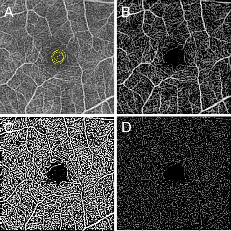
Retrospective, cross-sectional, observational study of healthy and diabetic adult subjects with and without DR. Retinal microvascular changes were assessed by using SD-OCTA images and an intensity-based optical microangiography algorithm. A semiautomated program was used to calculate indices of microvascular density and morphology in nonsegmented and segmented SD-OCTA images. Microvascular density was quantified by using skeleton density (SD) and vessel density (VD), while vessel morphology was quantified as fractal dimension (FD) and vessel diameter index (VDI). Statistical analyses were performed by using the Student's t-test or analysis of variance with post hoc Tukey honest significant difference tests for multiple comparisons.

RESULTS

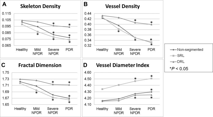
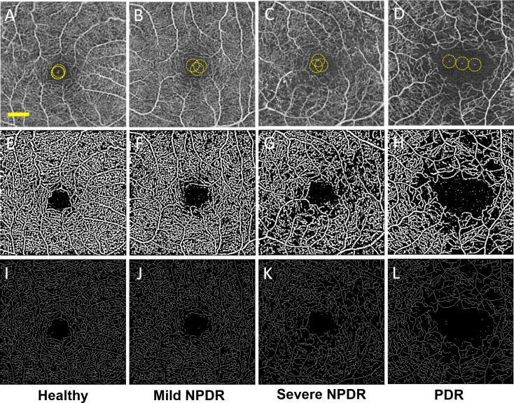
Eighty-four eyes with DR and 14 healthy eyes were studied. Spearman's rank test demonstrated a negative correlation between DR severity and SD, VD, and FD, and a positive correlation with VDI (ρ = -0.767, -0.7166, -0.768, and +0.5051, respectively; P < 0.0001). All parameters showed high reproducibility between graders (ICC = 0.971, 0.962, 0.937, and 0.994 for SD, VD, FD, and VDI, respectively). Repeatability (κ) was greater than 0.99 for SD, VD, FD, and VDI.

CONCLUSIONS

Vascular changes in DR can be objectively and reliably characterized with SD, VD, FD, and VDI. In general, decreasing capillary density (SD and VD), branching complexity (FD), and increasing average vascular caliber (VDI) were associated with worsening DR. Changes in capillary density and morphology were significantly correlated with diabetic macular edema.

摘要

目的

通过使用谱域光学相干断层扫描血管造影(SD-OCTA)来量化糖尿病视网膜病变(DR)中视网膜微血管的变化。

方法

对患有和未患有DR的健康及糖尿病成年受试者进行回顾性、横断面观察性研究。使用SD-OCTA图像和基于强度的光学微血管造影算法评估视网膜微血管变化。使用半自动程序计算非分割和分割的SD-OCTA图像中的微血管密度和形态学指标。微血管密度通过骨架密度(SD)和血管密度(VD)进行量化,而血管形态通过分形维数(FD)和血管直径指数(VDI)进行量化。使用Student's t检验或方差分析以及事后Tukey诚实显著差异检验进行多重比较的统计分析。

结果

研究了84只患有DR的眼睛和14只健康眼睛。Spearman秩检验显示DR严重程度与SD、VD和FD之间呈负相关,与VDI呈正相关(分别为ρ = -0.767、-0.7166、-0.768和 +0.5051;P < 0.0001)。所有参数在分级者之间均显示出高重现性(SD、VD、FD和VDI的组内相关系数分别为0.971、0.962、0.937和0.994)。SD、VD、FD和VDI的重复性(κ)大于0.99。

结论

DR中的血管变化可以通过SD、VD、FD和VDI进行客观可靠的表征。一般来说,毛细血管密度(SD和VD)降低、分支复杂性(FD)降低以及平均血管口径(VDI)增加与DR恶化相关。毛细血管密度和形态的变化与糖尿病性黄斑水肿显著相关。

https://cdn.ncbi.nlm.nih.gov/pmc/blobs/e535/4968771/7b8ec8aec557/i1552-5783-57-9-OCT362-f03.jpg
https://cdn.ncbi.nlm.nih.gov/pmc/blobs/e535/4968771/2fe30d5ed412/i1552-5783-57-9-OCT362-f01.jpg
https://cdn.ncbi.nlm.nih.gov/pmc/blobs/e535/4968771/453c06cc13e3/i1552-5783-57-9-OCT362-f02.jpg
https://cdn.ncbi.nlm.nih.gov/pmc/blobs/e535/4968771/7b8ec8aec557/i1552-5783-57-9-OCT362-f03.jpg
https://cdn.ncbi.nlm.nih.gov/pmc/blobs/e535/4968771/2fe30d5ed412/i1552-5783-57-9-OCT362-f01.jpg
https://cdn.ncbi.nlm.nih.gov/pmc/blobs/e535/4968771/453c06cc13e3/i1552-5783-57-9-OCT362-f02.jpg
https://cdn.ncbi.nlm.nih.gov/pmc/blobs/e535/4968771/7b8ec8aec557/i1552-5783-57-9-OCT362-f03.jpg

相似文献

1
Quantifying Microvascular Density and Morphology in Diabetic Retinopathy Using Spectral-Domain Optical Coherence Tomography Angiography.使用频域光学相干断层扫描血管造影术量化糖尿病视网膜病变中的微血管密度和形态。
Invest Ophthalmol Vis Sci. 2016 Jul 1;57(9):OCT362-70. doi: 10.1167/iovs.15-18904.
2
Quantifying Retinal Microvascular Changes in Uveitis Using Spectral-Domain Optical Coherence Tomography Angiography.使用频域光学相干断层扫描血管造影术量化葡萄膜炎中的视网膜微血管变化。
Am J Ophthalmol. 2016 Nov;171:101-112. doi: 10.1016/j.ajo.2016.08.035. Epub 2016 Sep 2.
3
Quantitative microvascular analysis of retinal venous occlusions by spectral domain optical coherence tomography angiography.基于频域光学相干断层扫描血管造影术的视网膜静脉阻塞微血管定量分析
PLoS One. 2017 Apr 24;12(4):e0176404. doi: 10.1371/journal.pone.0176404. eCollection 2017.
4
Interaction Between the Distribution of Diabetic Retinopathy Lesions and the Association of Optical Coherence Tomography Angiography Scans With Diabetic Retinopathy Severity.糖尿病视网膜病变病变分布与光学相干断层扫描血管造影扫描与糖尿病视网膜病变严重程度的相关性分析。
JAMA Ophthalmol. 2020 Dec 1;138(12):1291-1297. doi: 10.1001/jamaophthalmol.2020.4516.
5
Non-invasive structural and metabolic retinal markers of disease activity in non-proliferative diabetic retinopathy.非增生性糖尿病性视网膜病变中疾病活动的无创性结构和代谢性视网膜标志物。
Acta Ophthalmol. 2021 Nov;99(7):790-796. doi: 10.1111/aos.14761. Epub 2021 Jan 8.
6
Vessel Density around Foveal Avascular Zone as a Potential Imaging Biomarker for Detecting Preclinical Diabetic Retinopathy: An Optical Coherence Tomography Angiography Study.黄斑中心无血管区周围血管密度作为一种潜在的影像学生物标志物用于检测临床前期糖尿病视网膜病变: 一项光学相干断层扫描血管造影研究。
Semin Ophthalmol. 2020 Aug 17;35(5-6):316-323. doi: 10.1080/08820538.2020.1845386. Epub 2020 Dec 1.
7
Optical coherence tomography angiography analysis of the choriocapillary layer in treatment-naïve diabetic eyes.初诊糖尿病患者眼部脉络膜毛细血管层的光学相干断层扫描血管造影分析
Graefes Arch Clin Exp Ophthalmol. 2019 Jul;257(7):1393-1399. doi: 10.1007/s00417-019-04326-x. Epub 2019 May 14.
8
Macular vessels density in diabetic retinopathy: quantitative assessment using optical coherence tomography angiography.糖尿病视网膜病变中的黄斑血管密度:使用光学相干断层扫描血管造影术进行定量评估。
Int Ophthalmol. 2019 Aug;39(8):1845-1859. doi: 10.1007/s10792-018-1013-0. Epub 2018 Sep 7.
9
Optical Coherence Tomography Angiography Analysis of the Foveal Avascular Zone and Macular Vessel Density After Anti-VEGF Therapy in Eyes With Diabetic Macular Edema and Retinal Vein Occlusion.糖尿病性黄斑水肿和视网膜静脉阻塞患者抗VEGF治疗后黄斑无血管区和黄斑血管密度的光学相干断层扫描血管造影分析
Invest Ophthalmol Vis Sci. 2017 Jan 1;58(1):30-34. doi: 10.1167/iovs.16-20579.
10
Intereye Microvascular Differences in Patients With Same-Stage Diabetic Retinopathy Revealed by OCTA.OCTA 揭示同阶段糖尿病视网膜病变患者的眼内微血管差异。
Invest Ophthalmol Vis Sci. 2024 Jun 3;65(6):11. doi: 10.1167/iovs.65.6.11.

引用本文的文献

1
Efficacy of Faricimab in the Treatment of Diabetic Macular Edema and Faricimab-Related Changes in OCT and OCT Angiography.法西单抗治疗糖尿病性黄斑水肿的疗效以及光学相干断层扫描(OCT)和OCT血管造影中与法西单抗相关的变化
Pharmaceutics. 2025 Jun 30;17(7):858. doi: 10.3390/pharmaceutics17070858.
2
Predictive Vascular Changes in OCTA in Diabetic Patients.糖尿病患者光学相干断层扫描血管造影中的预测性血管变化
Biomedicines. 2025 Jun 17;13(6):1486. doi: 10.3390/biomedicines13061486.
3
Diet, Mitochondrial Dysfunction, Vascular Endothelial Damage, and the Microbiome: Drivers of Ocular Degenerative and Inflammatory Diseases.

本文引用的文献

1
Automated Quantification of Capillary Nonperfusion Using Optical Coherence Tomography Angiography in Diabetic Retinopathy.利用光学相干断层扫描血管造影术自动定量分析糖尿病视网膜病变中的毛细血管无灌注情况。
JAMA Ophthalmol. 2016 Apr;134(4):367-73. doi: 10.1001/jamaophthalmol.2015.5658.
2
Minimizing projection artifacts for accurate presentation of choroidal neovascularization in OCT micro-angiography.在光学相干断层扫描微血管造影中最小化投影伪影以准确呈现脉络膜新生血管。
Biomed Opt Express. 2015 Sep 28;6(10):4130-43. doi: 10.1364/BOE.6.004130. eCollection 2015 Oct 1.
3
DETECTION OF MICROVASCULAR CHANGES IN EYES OF PATIENTS WITH DIABETES BUT NOT CLINICAL DIABETIC RETINOPATHY USING OPTICAL COHERENCE TOMOGRAPHY ANGIOGRAPHY.
饮食、线粒体功能障碍、血管内皮损伤与微生物群:眼部退行性和炎症性疾病的驱动因素
Ophthalmol Ther. 2025 May 28. doi: 10.1007/s40123-025-01160-9.
4
HD-OCT Angiography and SD-OCT in Patients with Mild or No Clinically Apparent Diabetic Retinopathy.轻度或无临床明显糖尿病视网膜病变患者的高清光学相干断层扫描血管造影和光谱域光学相干断层扫描
Biomedicines. 2025 May 20;13(5):1251. doi: 10.3390/biomedicines13051251.
5
Deep learning segmentation of periarterial and perivenous capillary-free zones in optical coherence tomography angiography.光学相干断层扫描血管造影术中动脉周围和静脉周围无毛细血管区的深度学习分割
J Biomed Opt. 2025 May;30(5):056005. doi: 10.1117/1.JBO.30.5.056005. Epub 2025 May 8.
6
The association between urinary caffeine and caffeine metabolites and diabetic retinopathy in individuals with diabetes: NHANES 2009-2014.2009 - 2014年美国国家健康与营养检查调查:糖尿病患者尿中咖啡因及其代谢物与糖尿病视网膜病变之间的关联
Sci Rep. 2025 May 6;15(1):15827. doi: 10.1038/s41598-025-01088-x.
7
Recent advances and applications of optical coherence tomography angiography in diabetic retinopathy.光学相干断层扫描血管造影在糖尿病视网膜病变中的最新进展与应用
Front Endocrinol (Lausanne). 2025 Apr 16;16:1438739. doi: 10.3389/fendo.2025.1438739. eCollection 2025.
8
Peripapillary Bruch's membrane Opening-Minimum Rim Width (BMO-MRW) and microvascular changes in early diabetic retinopathy.视乳头周围脉络膜毛细血管层开口-最小边缘宽度(BMO-MRW)与早期糖尿病视网膜病变中的微血管变化
BMC Ophthalmol. 2025 Apr 28;25(1):257. doi: 10.1186/s12886-025-04077-w.
9
Association of Retinal Microvasculature With Myopia Progression in Children: The Hong Kong Children Eye Study.儿童视网膜微血管与近视进展的关联:香港儿童眼研究
Invest Ophthalmol Vis Sci. 2025 Apr 1;66(4):64. doi: 10.1167/iovs.66.4.64.
10
Biomarkers of ocular manifestation in newly diagnosed giant cell arteritis.新诊断巨细胞动脉炎眼部表现的生物标志物
BMC Ophthalmol. 2025 Apr 14;25(1):201. doi: 10.1186/s12886-025-03997-x.
利用光学相干断层扫描血管造影术检测糖尿病患者但无临床糖尿病视网膜病变患者眼部的微血管变化
Retina. 2015 Nov;35(11):2364-70. doi: 10.1097/IAE.0000000000000882.
4
CAPILLARY PLEXUS ANOMALIES IN DIABETIC RETINOPATHY ON OPTICAL COHERENCE TOMOGRAPHY ANGIOGRAPHY.光学相干断层扫描血管造影显示糖尿病视网膜病变中的毛细血管丛异常
Retina. 2015 Nov;35(11):2384-91. doi: 10.1097/IAE.0000000000000859.
5
RETINAL VASCULAR PERFUSION DENSITY MAPPING USING OPTICAL COHERENCE TOMOGRAPHY ANGIOGRAPHY IN NORMALS AND DIABETIC RETINOPATHY PATIENTS.使用光学相干断层扫描血管造影术对正常人和糖尿病视网膜病变患者进行视网膜血管灌注密度映射
Retina. 2015 Nov;35(11):2353-63. doi: 10.1097/IAE.0000000000000862.
6
ENLARGEMENT OF FOVEAL AVASCULAR ZONE IN DIABETIC EYES EVALUATED BY EN FACE OPTICAL COHERENCE TOMOGRAPHY ANGIOGRAPHY.通过正面光学相干断层扫描血管造影评估糖尿病患者眼部黄斑血管区扩大情况。
Retina. 2015 Nov;35(11):2377-83. doi: 10.1097/IAE.0000000000000849.
7
OPTICAL COHERENCE TOMOGRAPHY ANGIOGRAPHY OF RETINAL VENOUS OCCLUSION.视网膜静脉阻塞的光学相干断层扫描血管造影
Retina. 2015 Nov;35(11):2323-31. doi: 10.1097/IAE.0000000000000811.
8
Optical Coherence Tomography Angiography of Diabetic Retinopathy in Human Subjects.人体糖尿病视网膜病变的光学相干断层扫描血管造影
Ophthalmic Surg Lasers Imaging Retina. 2015 Sep;46(8):796-805. doi: 10.3928/23258160-20150909-03.
9
A morphological study of the foveal avascular zone in patients with diabetes mellitus using optical coherence tomography angiography.利用光学相干断层扫描血管造影术对糖尿病患者黄斑无血管区进行的形态学研究。
Graefes Arch Clin Exp Ophthalmol. 2016 May;254(5):873-9. doi: 10.1007/s00417-015-3143-7. Epub 2015 Sep 7.
10
Optical coherence tomography angiography of the foveal avascular zone in diabetic retinopathy.糖尿病视网膜病变中黄斑无血管区的光学相干断层扫描血管造影
Graefes Arch Clin Exp Ophthalmol. 2016 Jun;254(6):1051-8. doi: 10.1007/s00417-015-3148-2. Epub 2015 Sep 4.