Department of Neurology, Cognition and Aging Center, Kaohsiung Chang Gung Memorial Hospital, Chang Gung University College of Medicine, Kaohsiung, Taiwan.
Department of Radiology, Kaohsiung Chang Gung Memorial Hospital, Chang Gung University College of Medicine, Kaohsiung, Taiwan.
Sci Rep. 2018 Jan 24;8(1):1541. doi: 10.1038/s41598-018-19387-x.
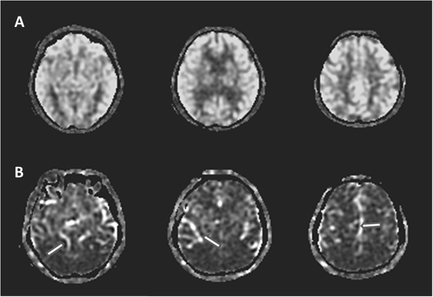
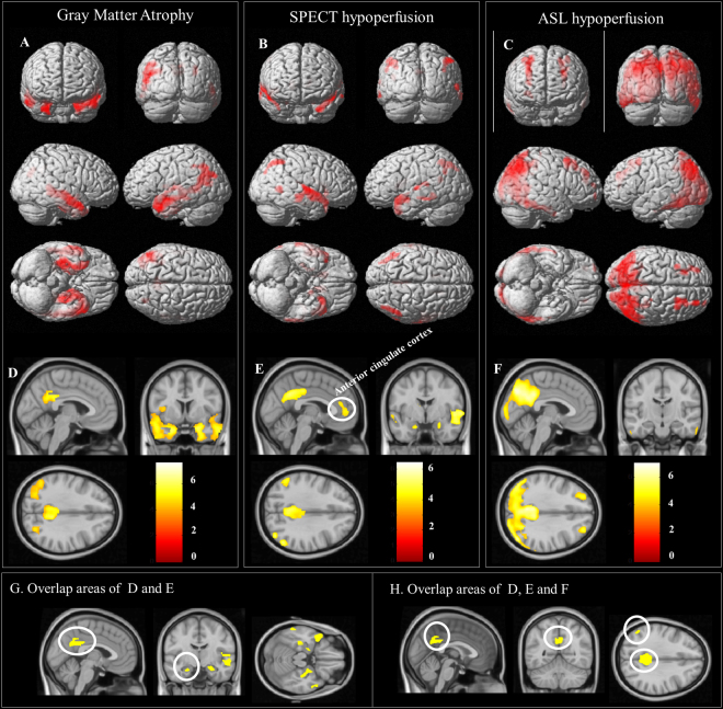
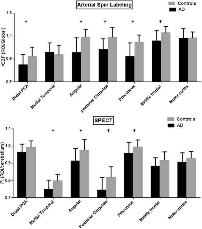
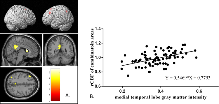
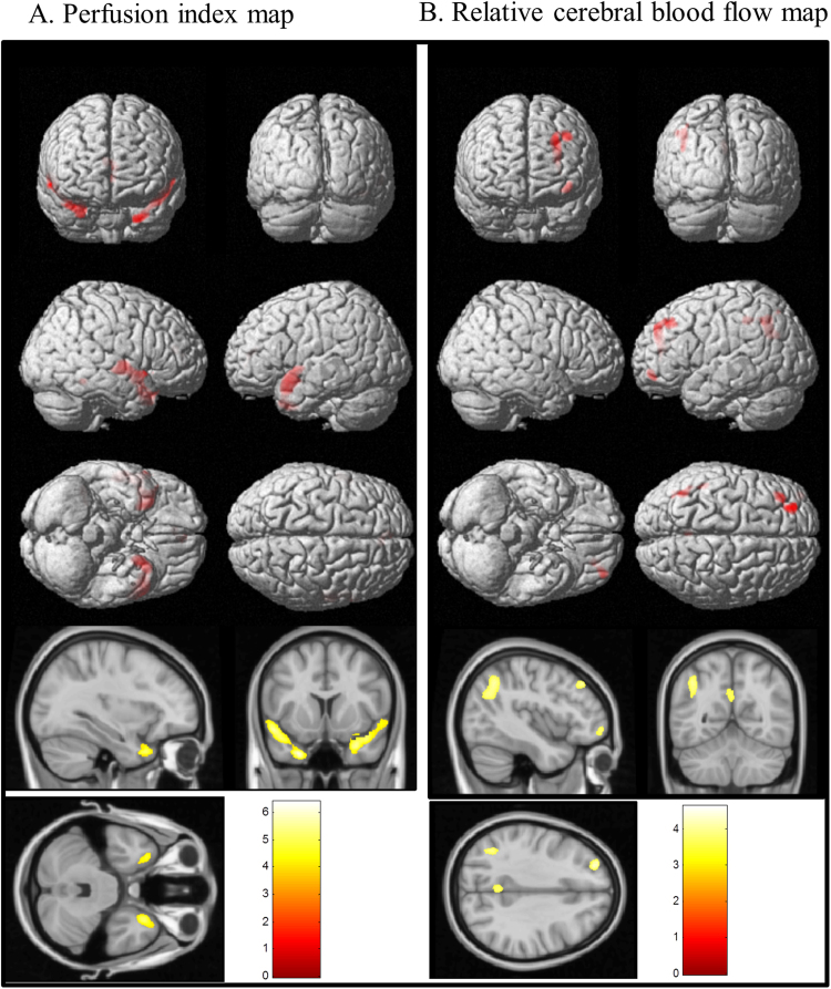
Micro- or macro-circulatory insufficiency has a negative impact in patients with Alzheimer's disease (AD). This study used arterial spin-labeled magnetic resonance imaging (ASL-MRI) and ethylcysteinate dimer single-photon emission computed tomography (ECD-SPECT) in 50 patients with AD and 30 age-matched controls to investigate how hypoperfusion patterns were associated with gray matter atrophy and clinical data. All participants completed 3DT1-MRI, ECD-SPECT and ASL-MRI examinations. Medial temporal cortex (MTC) volumes were correlated with regional signals showing significantly lower relative cerebral blood flow (rCBF) in ASL-MRI or perfusion index (PI) in ECD-SPECT. Neurobehavioral scores served as the outcome measures. Regions with lower PI showed spatial similarities with atrophy in the medial, anterior and superior temporal lobes, posterior cingulate cortex and angular gyrus, while regions showing lower rCBF were localized to the distal branches of posterior cerebral artery territories (posterior parietal and inferior temporal lobe) and watershed areas (angular gyrus, precuneus, posterior cingulate gyrus and middle frontal cortex). rCBF values in watershed areas correlated with MTC volumes and language composite scores. Precuneus and angular gyrus hypoperfusion were associated with the corresponding cortical atrophy. Macro- or micro-vasculature perfusion integrities and cortical atrophy determined the overall perfusion imaging topography and contributed differently to the clinical outcomes.
微循环或微循环不足会对阿尔茨海默病(AD)患者产生负面影响。本研究使用动脉自旋标记磁共振成像(ASL-MRI)和乙-半胱氨酸二聚体单光子发射计算机断层扫描(ECD-SPECT)对 50 名 AD 患者和 30 名年龄匹配的对照者进行研究,以探讨低灌注模式如何与灰质萎缩和临床数据相关。所有参与者均完成了 3DT1-MRI、ECD-SPECT 和 ASL-MRI 检查。内侧颞叶皮质(MTC)体积与区域信号相关,ASL-MRI 中相对脑血流(rCBF)或 ECD-SPECT 中灌注指数(PI)明显较低的区域信号相关。神经行为评分作为结局指标。PI 较低的区域与内侧、前上颞叶、后扣带回和角回的萎缩具有空间相似性,而 rCBF 较低的区域定位于大脑后动脉区域(后顶叶和下颞叶)和分水岭区域(角回、楔前叶、后扣带回和中额叶皮质)的远侧分支。分水岭区域的 rCBF 值与 MTC 体积和语言综合评分相关。顶内回和角回的灌注不足与相应的皮质萎缩相关。宏观或微观血管灌注完整性和皮质萎缩决定了整体灌注成像的形态,并对临床结局产生不同的影响。